
Page 60 - 2018_11-Haematologica-web
P. 60
C. Ka et al.
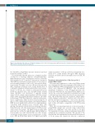
Figure 2. Liver histology of the index case of family 1 at diagnosis. Perls’ stain of liver biopsy shows significant amounts of stainable iron in Kupffer macrophages, while mild iron overload is observed in hepatocytes.
the SLC40A1 p.Arg178Gln missense mutation and had normal iron indices (3.III.1).
It is noteworthy that the index case of family 2 and his son had two co-existing conditions of hyperferritinemia: hemochromatosis type 4 and hepatic steatosis. Transient Elastography-based Controlled Attenuation Parameter (TE-CAP) measurements revealed grade 3 severe steatosis in both family members (CAP scores: 396 and 363 dB/m, respectively). The index case was a 55-year-old male with 36% transferrin saturation and a serum ferritin level of 1,452 mg/L at diagnosis. He presented with a waist circum- ference of 104 cm, in a context of subnormal laboratory metabolic and liver tests: uric acid (453 mol/L; normal range: 240-420), total cholesterol (7 mmol/L; normal range: 3.5-6.0), alanine aminotransferase (58 IU/L; normal range: 5-50) and gamma-glutamyl transferase (61 IU/L; normal range: 5-55). Blood pressure, aspartate aminotrans- ferase, triglycerides, HDL cholesterol and fasting blood glucose were within normal ranges. Abdominal magnetic MRI showed significantly reduced liver signal intensity, consistent with advanced iron overload (HIC: 180 μmol/g). The patient started venesection therapy when he turned 60 years old, after being diagnosed with prostate cancer and colon polyps. The phlebotomy program (500 mL every two weeks for eight months, then monthly for four months) was well tolerated. His son, who became overweight during infancy (waist circumference at diag- nosis: 105 cm; Body Mass Index: 31.7 kg/m2), presented
similar iron indices at the age of 20 years (transferrin satu- ration: 27%; serum ferritin: 976 mg/L). MRI, however, revealed a moderate increase in hepatic iron store (HIC: 85 mmol/g).
Functional characterization of the ferroportin 1 p.Arg178Gln variant
The functional significance of the p.Arg178Gln variant was determined by first investigating its subcellular local- ization. Wild-type (WT) and mutant ferroportin 1-V5 con- structs were expressed in HEK293T cells, and plasma membrane localization of the V5-tagged proteins was assayed by Western blot and densitometry. HLA-A was used as the control and standard for normalization, being a cell-surface protein with no known role in iron metabo- lism. The p.Ala77Asp missense mutation, which signifi- cantly damages ferroportin 1 structure and is known to prevent cell-surface localization,17,21 was used as the nega- tive control. The p.Arg178Gln mutant was properly local- ized on the cell surface, comparable to the WT protein (Figure3A).
Next, the iron-exporting function of the ferroportin 1 p.Arg178Gln variant was assessed using radioactively labeled iron. HEK293T cells were grown in 20 mg/L 55Fe-transferrin for 24 hr, washed, transiently transfected, and placed in a serum-free medium. The amount of 55Fe exported into the supernatant was measured after a period of 36 hr using cells transfected with the commercial
1800
haematologica | 2018; 103(11)
