
Page 58 - 2018_11-Haematologica-web
P. 58
C. Ka et al.
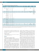
Table 1. Biological and clinical data of index cases and their relatives.
FamilyCountry Family Gender Age* SF TS HIC AST ALT GGT CRP RBC Hb Ht MCV Clinical Alcohol
1
2
3
relationships
(years) (mg/L) (%) ( mol/g) (IU/L) (IU/L) (IU/L) (mg/L)(Tera/L) (g/dL) (%) (fL) observations consumption**
Belgium Index case M Father M Daughter F Son M France Index case M Son M France Index case M Brother M Father M Grandmother F Uncle M First cousin F First cousin M 4 Belgium Indexcase M
39
61
11
7
55
20 976 27
15,7 45 Fatigue Moderate 15,2 42,5
12,6 Abstinent 12,9 Abstinent 15.6 45,8 96,5 Digestive problems Abstinent 18.4 52.1 90.9 Obesity Abstinent
Abstinent Abstinent
1873 33 1190
322 552
1452 36
180
85 60
37 58 61 1.5 4.75
54 98 23 2.3 5.73
12 6 35 72 45 21 22 54 29
773 33 382
1200
284
487
367
584 28
1114 26
Daughter F Son M Brother M Godson M
Goddaughter F
Iraq Index case M
France Index case M
Son M
reported by Taniguchi et al. in 201519 and, as templates, the exper- imental 3D structures of Bdellovibrio bacteriovorus (Bb) iron trans- porter Bd2019 in outward and inward facing conformations (pdb 5aym and 5ayo,19 respectively).
Statistical analysis
Data are presented as scatter dot plots and means. Comparisons used a one-tailed Student’s t-test.
Results
Clinical data and segregation analysis
The SLC40A1 p.Arg178Gln (c.533G>A) variant was identified in 22 patients from six independent families (Figure 1). It co-segregated with hyperferritinemia (defined as serum ferritin > 300 mg/L in males and > 200 mg/L in females) in all the tested patients, irrespective of age at testing, and was absent in three non-affected mem- bers of families 3 and 4.
Index cases comprised six males, aged 12-72 years at diagnosis, who displayed significant hyperferritinemia and non-elevated transferrin saturation (Table 1). Hepatic Iron Concentration (HIC) was evaluated using the Gandon’s magnetic resonance imaging (MRI) method20 in three patients with values between 60 (youngest case) and 250 mmol/g (oldest case) dry weight. Liver biopsy, performed in the index case of family 1, confirmed iron
263 21 25 613
5
6
59 1164 28 35 1354 36 32 372 25 71 2306 29 57 1386 43 27 1066 33
250
3830436
25112235
14,4 97 Fatigue Abstinent
15,4 45,3 91 Hepatomegaly Moderate
*Age at diagnosis. **Moderate: up to 1 drink per day for women and up to 2 drinks per day for men. SF: serum ferritin (normal value ≤ 200 μg/L in females, ≤ 300 in males); TS: transferrin sat- uration (normal value < 45%); HIC: hepatic iron concentration (normal value < 36 mmol/g); AST: aspartate aminotransferase (normal range: 5-50 IU/L); ALT: alanine aminotransferase (normal range: 5-50 IU/L); GGT: gamma-glutamyl transferase (normal range: 5-55 IU/L); CRP: C-reactive protein; RBC: red blood cell (normal range: 4.0-5.7 x 10¹²/L); Hb: hemoglobin (normal range: 12.0- 18.0 g/dL); Ht: hematocrit (normal range: 37-52%); MCV: mean corpuscular volume (normal range: 80-95 fL).
overload, with a predominance of iron deposition in Kupffer cells (Figure 2). The patient showed no clinical symptoms, except for persistent fatigue. The platelet count (178x109/L; normal range: 120-369x109/L) was not suggestive of a fibrotic liver disease. The patient started a phlebotomy program. After 24 phlebotomies (450 mL every three weeks), the hemoglobin level dropped to 13.6 g/dL, from 15.7 g/dL at diagnosis (normal range: 13.5 – 17.5).
In total, the cohort consisted of 17 males and five females. Women had lower serum ferritin levels than men (263-372 versus 487-2306 mg/L), while four children exhib- ited increased iron store between the ages of six and 12. Carrier testing in families identified 16 patients for whom no clinical manifestations of iron overload were reported.
Serum hepcidin measurements were obtained before therapeutic phlebotomy in two affected individuals of family 3 (3.II.5 and 3.III.4). They were above the reported range in healthy individuals for the assay used (1.0-21 ng/mL; liquid chromatography coupled with tandem mass spectrometry).16 The proband’s grandmother (3.I.1) start- ed therapeutic phlebotomy (1-2 venesections per year; well tolerated) several years before family screening. She showed moderately increased ferritin (280 μg/L) at the time of serum hepcidin evaluation (41.6 ng/mL). In con- trast, normal serum hepcidin levels (8.2 ng/mL) were detected in the proband’s brother who was negative for
1798
haematologica | 2018; 103(11)
