
Page 61 - 2018_11-Haematologica-web
P. 61
Ferroportin structure and function
A
B
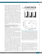
pcDNA3.1-V5-His vector as negative control (no FPN1). As shown in Figure 3B, cells transfected with WT ferro- portin 1-V5 displayed a 3-fold increase in iron release. The p.Arg178Gln variant was not able to export iron 55Fe in amounts comparable with WT ferroportin 1, but was more active than the p.Ala77Asp control; Student’s t-tests highlighted significant differences between both variants and WT ferroportin 1 (P<0.001 and 0.0001, respectively) and between the two variants (P<0.0001).
To investigate whether the p.Arg178Gln missense mutation could modify response to hepcidin, transiently transfected HEK293T cells were cultured for 24 hr with conditioned media derived from T-Rex-293 cells stably transfected with full-length human HAMP cDNA. Supernatant human-25 hepcidin concentration was deter- mined using a competitive enzyme-linked immunosor- bent assay. Two known ferroportin 1 mutants served as positive controls: p.Asn144His, which shows partial resistance to hepcidin inhibition,22 and p.Cys326Tyr, which abolishes hepcidin binding to ferroportin 1 and is responsible for complete resistance.23 As expected, the addition of hepcidin to cells expressing WT ferroportin 1 resulted in the disappearance of the iron exporter from the plasma membrane. The Western blot pattern of the p.Asp178Gln variant, unlike the p.Cys326Tyr and p.Asn144His mutants, was similar to that of the WT pro- tein (Figure 4).
Structural and functional investigation of the intermolecular interaction between the N and C lobes of human ferroportin 1 – 3D structure form I (outward facing)
In order to understand the possible impact of the p.Arg178Gln mutation, we modeled the 3D structure of human ferroportin in both the outward and inward facing conformations, based on the recent 3D structures of the Bb iron transporter Bd2019, which shares 24% sequence identity with human ferroportin 1.19
As illustrated in Figure 5, Arg178 forms an inter-lobe salt bridge with Asp473 in the outward facing conformation of human ferroportin 1. We hypothesized that this non- covalent interaction between helix TM5 and helix TM8, located respectively in the N and C lobes, might be impor- tant in stabilizing the outward facing conformation of fer- roportin 1, and that its disruption might cause a significant reduction in iron egress.
To check this hypothesis, we replaced arginine 178 and aspartic acid 473 with alanine, which is the smallest amino acid after glycine and is neutral, being non-polar and devoid of any strong hydrophobic character. Moreover, it has the highest propensity of the 20 amino acids for the a-helical state;24 thus this modification was likely to have limited impact on local structure. As shown in Figure 6, the Asp473Ala mutant did not cause obvious mislocalization of the protein, which was, however, total- ly inoperative for iron export. Indeed, cells expressing the Asp473Ala mutant retained 55Fe in amounts comparable to cells expressing the two known p.Asn77Asp and p.Val162del loss-of-function mutations. The Arg178Ala mutant reduced cell surface expression to half that of the WT, but with less influence in iron export ability.
We then examined whether charge swapping could restore the iron export function of ferroportin 1, and whether Arg178Gln dysfunction could be corrected by a p.Asp473Arg mutation. The p.Arg178Asp and
Figure 3. The ferroportin p.Arg178Gln mutant shows normal cell surface
expression, but substantial loss of iron export. (A) HEK293T cells were tran-
siently co-transfected with plasmids encoding either a V5-tagged ferroportin pro-
tein (WT or variant) or a V5-tagged HLA-A protein. Human leukocyte antigen
(HLA)-A was used as control and standard for normalization, being a cell surface
protein with no known role in iron metabolism. At 24h after transfection, cell sur-
face proteins were selectively purified and analyzed by Western blotting using a
peroxidase conjugated mouse anti-V5 antibody. Densitometric scans of
SLC40A1 levels (normalized to HLA-A) are shown in the lower part of the figure.
The results of three independent experiments are presented. (B) HEK293T cells
were grown in 20 mg/mL 55Fe-transferrin for 24h before being washed and tran-
siently transfected with WT or mutated SLC40A1-V5 expression plasmids. After
Fe exported into the supernatant was collected at 36h. Data are presented as percentage of cellular radioactivity at time zero. Each point represents the average value (from tripli- cate) of five independent experiments. P values were calculated with the
15h, cells were washed and then serum-starved. The Student’s t-test; **P<0.01 and ****P<0.0001. WT: wild-type.
55
p.Asp473Arg substitutions almost abolished cell surface expression of the protein. Introducing the two p.Arg178Asp and p.Asp473Arg missense mutations did not rescue the membrane expression level decreased by single mutations. The Arg178Gln/Asp473Arg double mutant also resulted in a strong reduction in cell surface expression (Online Supplementary Figure S1).
Taken together, these results suggested that the salt bridge between arginine 178 and aspartic acid 473 is essential for ferroportin 1 iron export function. Any subtle changes in charge or size on the side chains may cause loss of function.
haematologica | 2018; 103(11)
1801
