
Page 95 - Haematologica July
P. 95
CHD4 is required for maintenance of childhood acute myeloid leukemia
ABC
DE
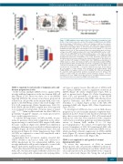
Figure 3. CHD4 inhibition causes minor effect in cell growth of normal blood cells in vitro. A. Bar charts represent real time PCR analysis of mRNA levels after shRNA- based knockdown of Chd4 relative to control cells transduced with vectors express- ing scrambled shRNA, in sorted primary murine cKit+ BMs used in the competitive proliferation assays (Figure 3B,C), at 72 hours post transduction. mRNA levels were normalized to Hprt. The data is represented as the mean ±S.E.M., ****P<0.001 (unpaired t-test), n=3. B. Flow cytometry charts are representative plots of the per- centage of live RFP+ Chd4 knockdown cKit+ cells (Chd4 shRNA-RFP) relative to GFP+ control cells (Sc shRNA-GFP) at the indicated time points. C. Line chart of a repre- sentative experiment of the percentage of Chd4 shRNA-RFP+ vs. Sc shRNA-GFP+ BMs propagated in suspension, normalized to 50% at the initial time point (Day one), determined by flow cytometry analysis at the indicated time points. D. Bar charts rep- resent real time PCR analysis of mRNA levels after shRNA-based knockdown of CHD4, relative to control cells transduced with vectors expressing scrambled shRNA, in primary human CD34 enriched UCBs used in the cell growth assays (Figure 3E), 72 hours post transduction. mRNA levels were normalized to UBC. The data is rep- resented as the mean ±S.E.M., ***P<0.005 (unpaired t-test), n=3. E. Bar chart of the total number of viable primary UCBs, transduced with shRNA against CHD4 (CHD4-shRNA), or a negative control vector (Sc-shRNA). The UCBs were propagated in suspension with supplemented cytokines and growth factors for 14 days. The number of viable UCBs was determined by flow cytometric analysis. **P<0.01 (unpaired t-test), n=3. mRNA: messenger ribonucleic acid; RFP: red fluorescent pro- tein; GFP: green fluorescent protein; shRNA: short hairpin RNA.
CHD4 is required for cell growth of leukemia cells and disease progression in vivo
In the screens, multiple shRNA vectors against CHD4 strongly inhibited expansion of the two human AML cell lines (Figure 1C,D). Moreover, three out of six shRNAs targeting CHD4 in the screens caused more pronounced reduction in cell growth in the murine AML cells com- pared to the FDCP-mix control cells (fold change: 0.19, 0.32, 0.38, respectively) (Online Supplementary Table S2). Both CHD4 and the NuRD complex have been reported to be required for cell growth of various types of cancer cells.9,13,14,37-39 Taken together with the fact that CHD4 is a potentially “druggable” enzyme, our studies proceeded to focus on this epigenetic factor.
To validate the importance of CHD4 in AML, we per- formed a pairwise cell growth competition assay, allow- ing monitoring of control and CHD4 targeted cells under the same conditions (Figure 2A). shRNA-based inhibition using two different vectors resulted in efficient reduction in mRNA (Figure 2B; Online Supplementary Figure S3A) and protein levels (Figure 2C; Online Supplementary Figure S3B) of CHD4 as compared to control cells transduced with a negative control vector.
Longitudinal flow cytometric analysis revealed that THP-1, NOMO-1 and mouse AML cells subjected to CHD4 knockdown by two independent shRNAs were strongly inhibited in cell growth compared to control cells (Figure 2D,E; Online Supplementary Figure S3C,D).
Given the demonstrated importance in MLL-rearranged AML cells, we then investigated whether CHD4 was also required for cell growth of leukemic cells carrying differ-
ent types of genetic lesions. Knockdown of CHD4 with two distinct shRNAs caused a significant reduction in mRNA (Figure 2F; Online Supplementary Figure S3E) as well as protein levels (Figure 2G; Online Supplementary Figure S3F), and prevented cell growth of the HL-60 (promyelocytic leukemia cells), K562 cells (chronic myel- ogenous leukemia) and NB-4 (acute promyelocytic leukemia), to a similar degree as that of the MLL-AF9 rearranged AML cells (Figure 2H,I; Online Supplementary Figure S3G,H).
To investigate if CHD4 also has a role in AML disease progression, we used a murine immune competent trans- plantation model.40 As previously reported, wild-type (wt) mice that received transplants of MLL-AF9 transformed AML mouse cells, that were transduced with a negative control vector, resulted in pathologic and clinical manifes- tations of human AML within 19 to 21 days post transplantation.40 In contrast, wt recipient mice that received transplants of MLL-AF9 transformed AML cells that were transduced with two individual shRNA vectors, which mediated efficient suppression in CHD4 mRNA levels (Figure 2J), survived significantly longer than mice transplanted with control cells (Figure 2K).
CDH4 is not required for cell growth of normal hematopoietic cells
To assess the importance of Chd4 in normal hematopoietic cells, we performed shRNA-based knock- down experiments in primary mouse BMs, which result- ed in efficient reduction in Chd4 mRNA levels compared to the control cells (Figure 3A). Using the cell growth
haematologica | 2018; 103(7)
1173
