
Page 53 - Haematologica July
P. 53
Mouse model of sickle cell disease
underlying these effects requires further validation in SCD mouse models, our study suggests that ETA receptors are key players in SCD-associated pain.
The present study used two humanized mouse models of SCD, the Berkeley and Townes, to analyze SCD-asso- ciated pain. Although pain-like behaviors in SCD mouse models may not exactly mimic pain in SCD patients due to several confounding environmental and emotional vari- ables, analyzing pain hypersensitivity in SCD murine models is a productive tool for identifying the underlying mechanisms of SCD-associated pain. These mice dis- played significant mechanical allodynia, thermal/cold hyperalgesia, and spontaneous ongoing pain consistent with previous reports.36 Using age-matched male and female HbSS and HbAA littermates, we found no signifi- cant sex-based differences in basal or post-hypoxia evoked pain hypersensitivity or ABT-627 efficacy. This aligns with clinical reports that found no significant differ- ences in self-reported pain experiences between maleand female SCD patients.37,38 Interestingly, Townes HbSS mice did not display a similar degree of pain exacerbation fol- lowing hypoxia/reoxygenation compared to the BerkSS mice. This may be related to the presence of a human β-globin locus control region (LCR) that leads to hemoglo- bin switching similar to humans in the Townes HbSS mice, although both models express human globins. In the Townes model, relative g-globin expression (g/g+βS) at birth is between 30% to 50%, then β-globin expression dominates at 1 month of age.39 In contrast, the Berkeley model exhibits a complete g-globin to β-globin switch in utero leading to greater prenatal death and low birth rates. Given that higher g-globin expression is associated with diminished SCD symptoms,40 the early expression of βs- globin in BerkSS mice may cause more severe symptoms of SCD, which could explain their significant pain exacer- bation following hypoxia/reoxygenation.
An increase in ETA receptor expression may be attrib- uted to the elevated ET-1 in the DRG neurons of HbSS
mice. The levels of ET-1 mRNA, ET-1 protein, and ETA receptor protein increased in HbSS DRG. Consistent with previous studies,10,25 ET-1 and ETA receptors are predomi- nantly expressed in DRG neurons, although ET-1 expres- sion in immune cells found in HbSS DRG cannot be ruled out.41 Elevated ET-1 could trigger increased ETA receptor expression in DRG possibly through increased receptor cycling to the membrane and/or enhanced transcription or translation.42,43 Since we found no significant changes in ETA receptor mRNA in HbSS DRG, it would be expected that, under constantly elevated ET-1 conditions, ET-1/ETA receptor cycling coupled with ET-1-triggered activation of intracellular signals enhances ETA receptor translation through unknown mechanisms in HbSS DRG. The mech- anism of ETA receptor upregulation in HbSS DRG remains to be further investigated.
ETA receptors are required for increased Nav1.8 expres- sion in HbSS DRG, since ETA receptor inhibition com- pletely blocked increases in Nav1.8 protein and current in HbSS DRG. However, whether the participation of DRG ETA receptors in the pain hypersensitivity of HbSS mice is mediated by DRG Nav1.8 upregulation in HbSS mice requires further investigation as the present data showed only an association between activation of the ETA recep- tors and upregulation of Nav1.8 in the DRG of HbSS mice. Interestingly, ET receptor inhibition did not alleviate the
channels in HbSS DRG. Previous studies reported that
phosphorylation of Nav1.8 channels by protein kinase A
(PKA), but not PKC, could affect the channel’s gating prop-
erties.44,45 The altered gating of Nav1.8 channels in the
HbSS DRG is likely the result of activation of PKA by an
ET receptor-independent signaling mechanism. It is A
worth noting that the leftward shift of 4.44 mV in the HbSS DRG would only result in a modest increase in sodi- um current.26 Given that ABT-627 fully attenuated the increase in Nav1.8 current in the HbSS DRG, an ETA -receptor independent mechanism of Nav1.8 channel
A
hyperpolarizing shift in the gating properties of Nav1.8
A
B
CD
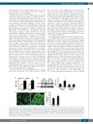
Figure 6. Nav1.8 expression is elevated in Townes HbSS DRG and is ETA receptor-dependent. (A) Expression of Scn10a mRNA (encoding Nav1.8) and Scn11a mRNA (encoding Nav1.9) in the L3-L4 DRG from HbAA mice and HbSS mice. n=3 mice/genotype. *P<0.05 vs. the corresponding HbAA mice. (B) Effect of hindpaw injection of ABT-627 or vehicle on expression of Nav1.8 protein in the L3-L4 DRG from HbAA mice and HbSS mice. Left: representative Western blots. Right: a summary of densitometric analysis. n=3 mice/group. *P<0.05 vs. the vehicle-treated HbAA mice. (C) Number of Nav1.8-labeled neurons in the L4 DRG from HbAA mice and HbSS mice. Left: representative immunostaining images. Right: a summary of statistical analysis of the number of Nav1.8-labeled neurons. Scale bar: 50 mm. n = 3 mice/genotype. **P<0.01 vs. the HbAA mice.
haematologica | 2018; 103(7)
1131
