
Page 65 - Haematologica June
P. 65
engraftment, we further compared engraftment of MDS mononuclear cells without MSC to healthy donor-derived MSC (normal), or allogeneic patient-derived MSC (allo) and to patient-derived autologous MSC (auto) (Figure 3A). Although some mice showed increased engraftment with different MSC samples tested, no consistent pattern of enhanced engraftment was seen that correlated with the source of the MSC (Figure 5E).
To better understand the transient effect of the presence of MSC on the levels of engraftment observed for some of the patients’ samples tested, in vitro-expanded MSC were labeled using a lentiviral plasmid expressing green fluores- cent protein and luciferase. Those cells were further trans- planted via intrabone injections in NSG mice and moni- tored over time. In vivo imaging revealed gradually decreasing levels of luminescence (Figure 5F). Luminescence was solely detected in the area of the inject- ed femur and its levels were completely diminished by week 4 after transplantation.
Long-term engraftment of myelodysplastic syndrome cells in NSG-S mice
To evaluate whether long-term engraftment of MDS cells can be achieved in NSG-S mice, human CD45 cells, isolated from the bone marrow of well-engrafted primary recipients, were selected and intrafemorally transplanted
into secondary recipients with or without MSC according to the experimental plan (Figure 6A). Thirteen weeks after transplantation the mice were sacrificed. Analysis of the different subpopulations identified the presence of myeloid CD33+ cells, as well as CD34+CD38- cells. As in the primary recipients, no B or T cells were detected (Figure 6B). Small erythroid populations in secondary recipients were also unable to differentiate further to more mature erythroid cells (Figure 6C). All mice showed increased levels of engraftment compared to the engraft- ment levels of the primary recipients at the same time
Table 2. Clinical samples used for the MDS engraftment studies.
Cytokines increase AML but not MDS engraftment
Patient ID#
2970
2381 108 3598 2952 4712 3282
Diagnosis
MDS/MPN
Therapy related myeloid neoplasm Unclassified MDS MDS-EB-1
MDS-EB-1
MDS-EB-1
MDS-EB-2
Risk level
Low
Low Low High High High High
MDS: myelodysplastic syndrome; MPN: myeloproliferative neoplasm; EB: excess blasts.
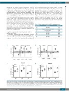
AB
CD
Figure 4. Most myeloproliferative syndrome (MDS) samples do not show sustained engraftment and human cytokines moderately enhance engraftment. Time course representation of the percentages of hCD45+ cells in the bone marrow of xenografted NSG-S mice injected with (A) high risk and (B) low risk human MDS patients’ mononu- clear cells. The gray line corresponds to the 0.1% threshold used in the study and the gray zone reflects a broader area of uncertainty of engraftment. Bone marrow mononuclear cells from patients diagnosed with MDS were intrafemorally injected into NSG and NSG-S mice in a strain comparison experiment. Percentage of hCD45+ cells found in the murine bone marrow was evaluated at (C) eight and (D) 16 weeks after transplantation.
haematologica | 2018; 103(6)
967
