
Page 63 - Haematologica June
P. 63
D
E
B
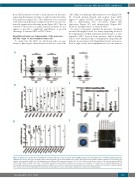
Figure 2. NSG-S mice enhanced the engraftment of human patients’ acute myeloid leukemia (AML) samples more than NSG mice (non-NSG engrafted patients’ samples were used). (A) Levels of hCD45+CD33+ leukemia blasts in bone marrow (BM), spleen (SPL) and peripheral blood (PB) of NSG or NSG-S mice (n=14) injected with the same number of AML cells. (B) Individual mouse BM and SPL leukemia burdens of 11 out of the 14 mice from (A) are shown. (C) Receptor densities (CD116, CD117 and CD123) on AML cells of the 7 non-NSG-S engrafters and 14 NSG-S engrafters were assessed as number of receptors per cell. (D) Establishment of 8 inv(16) AML patient-derived xenotransplant models in NSG-S mice. (E) The chromosomal abnormalities for inv(16) were confirmed in the BM of engrafted NSG-S mice by fluorescent in situ hybridization (left panels) and breakpoint reverse transcriptase polymerase chain reaction (right panel).
Cytokines increase AML but not MDS engraftment
from MDS patients revealed a small increase in the mice expressing the human cytokines at eight weeks after intra- bone injections (Figure 4C). This difference was sustained at 16 weeks in all patients’ samples tested but was not sta- tistically significant at this time point (Figure 4D). Thus, in contrast to their effect on AML, human cytokines do not appear to provide a significant engraftment or growth advantage to human MDS in NSG-S mice.
Engraftment levels are independent of the presence and the origin of mesenchymal stem cells
To verify that the MSC are functional cells, we per- formed a phenotypic characterization and also tested the
A
cells’ ability for trilineage differentiation in vitro (Figure 5A- D). Overall, patient-derived and normal donor MSC appear to express all MSC markers (Figure 5A) and are able to differentiate into osteoblasts (Figure 5B), adipocytes (Figure 5C) and chondrocytes (Figure 5D), therefore having features of bona fide MSC.
In order to investigate whether the presence of human stromal cells might be used as a better supporting tissue for the engraftment of MDS in immunodeficient mice, in vitro- expanded MSC derived from patients and/or healthy donors were intrafemorally co-transplanted along with the patients’ mononuclear cells. Engraftment levels were meas- ured at eight weeks after transplantation by bone marrow
C
haematologica | 2018; 103(6)
965
