
Page 61 - Haematologica June
P. 61
Cytokines increase AML but not MDS engraftment
continued from the previous page
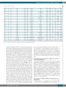
3918 73 M M5 2258 57 M M5 2698 43 F M5 2844 73 M M5 4090 68 M NA 2750 72 M NA
53 59 F NA 3196 64 M NA 3365 62 M NA 3949 59 F NA 4133 58 M NA 2339 47 M ND 3055 54 F NOS 2748 64 F NOS 1932 31 F NOS 2933 56 M NOS 3081 59 M NOS 3033 74 M NOS
774 37 F NOS 2741 52 M NOS 2837 43 F NOS 3261 39 M NOS 1897 64 F Secondary
0.1397 PB 0.0429 PB 0.1298 PB 0.3246 PB 0.0979 PB 0.0983 PB 0.1836 PB 0.0711 PB 0.0839 PB 0.0778 PB
De novo De novo De novo De novo NA
De novo
NA Relapsed De novo Refractory Relapsed Relapsed De novo De novo Relapsed De novo De novo
Normal WT NA NA
MUT 40
NA 77 NANND
NAN MSC
NA PB
0.349 PB
0.431 PB
0.1026 PB
0.215 PB
0.0476 PB
0.0224 BM
0.038 BM Denovo
ITD MUT 87
WT WT 51
NA NA 25
WT WT 73
ITD NA NA
ITD NA 87
ITD MUT 22
ITD NA 94
ITD NA 72
WT NA 80
ITD MUT NA
NA MUT 97
NA NA NA
WT WT 76
WT MUT 79
WT WT NA
ITD NA NA
ND NA NA
ITD WT 91
ITD MUT NA
WT NA NA
WT NA 94
WT NA 90 NANND
NA 45,X,-MSC/46,XY ND
Normal Normal 47,XY,+8, t(3;17) Normal Normal
NA
NA
Normal Normal Normal Normal Normal
NA t(7;11)(p15;p15) Complex 47,XX,+8 Normal t(8;16)(p11.2;p13.3)
NA MSC MSC 80 MSC MSC NAN MSC NA N N 70 MSC ND NA MSC ND 80 MSC ND 94 MSC MSC NA MSC MSC NAN ND NA MSC ND NA N N NA N ND NA N ND 90 N ND 68 N ND NA MSC ND NA MSC MSC NA MSC MSC NA MSC MSC 90 N MSC NA MSC ND
0.1245 PB De novo
0.1317 0.0864 0.0955 0.0348
PB Relapsed
De novo
De novo
PB
PB
BM
BM Relapsed t(8;16)(p11.2;p13.3) PB Treatment related Complex
2107 64 F
2074 78 F
Secondary 0.0649
Secondary 0.0237
Treatment related
M: male; F: female; MSC: engrafted; N: not engrafted; ND: not done; NA: not available; PB: peripheral blood; BM: bone marrow; FAB: French-American-British classification; WBC: white blood cell count (x 109 cells/L); ITD:internal tandem duplication; WT: wild-type; MUT: mutated.
myeloid neoplasms) (Table 2). We performed intrabone injections directly into the femoral cavity at the orthotopic site as it has been described in patient-derived xenotrans- plantation models for AML, that intrabone cell transplan- tation results in a higher probability of successful engraft- ment. This can be advantageous for patient-derived xeno- transplantation models of MDS in particular, as the num- bers of bone marrow mononuclear cells are commonly modest or low.28 In order to address whether the presence of human stromal cells results in increased engraftment levels, MSC were ex vivo-expanded and co-injected along with the patients’ mononuclear cells. Levels of engraft- ment were assessed by bone marrow aspiration at differ- ent time points throughout the experiment and are expressed as the percentage of human CD45+ cells. To assess the subpopulations of cells engrafted, cells were fur- ther analyzed with the lineage-specific antibodies CD19 for B cells and CD3 for T cells as well as for the presence of stem and progenitor cells using the markers CD34 and CD38, CD123 and CD45RA, respectively (Figure 3B). In the majority of engrafted patients’ samples, B and T cells were not detected or were detected at uncommonly low levels at the time points tested. In contrast, the myeloid CD33+ component was present in all mice tested as well as subpopulations of CD34+ and CD38+ human cells. The ability to differentiate into cells of the erythroid lineage was assessed in the low and negative fractions of human CD45+ cells using the erythroid differentiation markers CD71 and glycophorin A. Even though erythroid cells at the initial stages of differentiation were detected in all mice tested, cells lacked the ability to differentiate further
into more mature cells (Figure 3C). These results were fur- ther confirmed by immunohistochemical analysis of decalcified bone sections, showing a broad presence of human CD33-stained cells, but absence of megakary- ocytes expressing GPIIIa and erythroid cells expressing glycophorin C (Figure 3D). Overall, these results confirm that MDS cells with multi-lineage potential can potential- ly be transferred and maintained in immunocompromised mice.
Most myelodysplastic syndrome samples do not show sustained engraftment
The term “engraftment” used in the context of xeno- transplantation studies is equated with long-term mainte- nance and, typically, expansion of transplanted cells. To quantitatively assess whether MDS cells engraft in NSG-S mice, we divided our samples into low- and high-risk MDS. As shown in Figure 4, engraftment of high-risk MDS was heterogeneous with 2 samples never showing robust engraftment but 2 samples showing early and sus- tained engraftment (Figure 4A). Only one of the 3 low-risk MDS samples demonstrated clear engraftment, whereas levels of engraftment decreased over time for the other 2 samples (Figure 4B). For comparison, a secondary AML sample shows the expected low early engraftment with increase over time (Figure 4A).
The presence of human cytokines marginally improves engraftment
Direct comparison of engraftment levels between NSG and NSG-S mice transplanted with mononuclear cells
haematologica | 2018; 103(6)
963
