
Page 214 - Haematologica May 2022
P. 214
Case Reports
Kikuchi-Fujimoto disease associated with hemo- phagocytic lymphohistiocytosis following the BNT162b2 mRNA COVID-19 vaccination
Kikuchi-Fujimoto disease (KD) is a self-limiting histio- cytic necrotizing lymphadenitis. One case of KD follow- ing administration of the BNT162b2 mRNA COVID-19 vaccine was recently reported.1 Hemophagocytic lym- phohistiocytosis (HLH) is a life-threatening hyperinflam- matory state brought on by uncontrolled histiocytes, macrophages and T-cell activation, which have also been occasionally observed after BNT162b2 vaccination.2 KD and HLH present overlapping pathogenesis and symp- toms, and their association has been previously described in children and adult patients.3,4 Here, we report the first case of KD associated with HLH following the BNT162b2 mRNA COVID-19 vaccination.
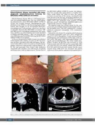
A 38-year-old previously healthy woman was admitted to the hospital with a history of a fever of 40°C for more than ten days, associated with chills and fatigue. She pre- sented with a diffuse cutaneous eruption of erythematous papules, which were subsequently confluent (Figure 1, A- B). She had previously been treated with antibiotics (amoxicillin and clavulanic acid, followed by teicoplanin and doxycycline), with no improvement. The first dose of
the BNT162b2 mRNA COVID-19 vaccine was adminis- tered two months after giving birth to a healthy baby. Three weeks before the onset of fever, the patient received a second dose of the vaccine, inoculated in the same left arm as the first dose. At hospital admission, her nasopharyngeal swab for SARS CoV-2 PCR was negative; SARS CoV-2 IgG antibodies were positive (>2.080 BAU/mL; cut-off: 33.8 BAU/mL; LIAISON SARS-CoV-2 TrimericS IgG, Diasorin, Saluggia, Italy).
Her physical exam was normal except for the cuta- neous rash and multiple enlarged tender lymph nodes in the left axillary zone. This was confirmed by a contrast- enhanced computed tomography (CT) examination (Figure 1, C-D).
Laboratory tests showed bi-cytopenia with leukopenia and anemia (neutrophil count, 0.9 x 10⁹/L; lymphocyte count, 0.3 x 10⁹/L, hemoglobin, 9.8 g/L), increased lactate dehydrogenase and transaminase levels, high serum fer- ritin levels (500 μg/L), mild hypertriglyceridemia (225 mg/dL) and normal fibrinogen. Her serum soluble inter- leukin-2 receptor (IL-2R) level was increased to 2.610 U/mL (normal value 223-710) and her natural killer (NK) cell count was low (<35 cells/μL; normal value 200-400). IL-6, IL-8 and IL-10 levels were normal, but the tumor necrosis factor (TNF) level was increased. The patient underwent a bone marrow aspiration and trephine proce-
AB
CD
Figure 1. Patient’s clinical and radiological findings. A and B) Diffuse maculo-papular skin rash; C and D) CT scan showing left axillary lymph node enlargement.
1222
haematologica | 2022; 107(5)
