
Page 215 - Haematologica May 2022
P. 215
Case Reports
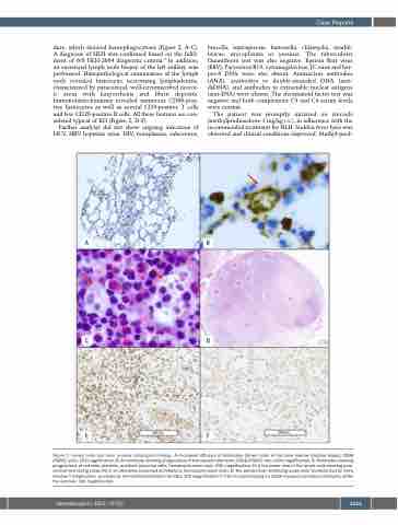
dure, which showed hemophagocytosis (Figure 2, A-C). A diagnosis of HLH was confirmed based on the fulfil- ment of 6/8 HLH-2004 diagnostic criteria.5 In addition, an excisional lymph node biopsy of the left axillary was performed. Histopathological examination of the lymph node revealed histiocytic necrotizing lymphadenitis, characterized by paracortical, well-circumscribed necrot- ic areas with karyorrhexis and fibrin deposits. Immunohistochemistry revealed numerous CD68-posi- tive histiocytes as well as several CD3-positive T cells and few CD20-positive B cells. All these features are con- sidered typical of KD (Figure 2, D-F).
Further analysis did not show ongoing infections of HCV, HBV hepatitis virus, HIV, toxoplasma, rubeovirus,
brucella, leptospirosis, bartonella, chlamydia, morbil- livirus, mycoplasma or yersinia. The tuberculosis Quantiferon test was also negative. Epstein Barr virus (EBV), Parvovirus B19, cytomegalovirus, JC virus and her- pes-6 DNA were also absent. Antinuclear antibodies (ANA), antibodies to double-stranded DNA (anti- dsDNA), and antibodies to extractable nuclear antigens (anti-ENA) were absent. The rheumatoid factor test was negative and both complement C3 and C4 serum levels were normal.
The patient was promptly initiated on steroids (methylprednisolone 1 mg/kg i.v.), in adherence with the recommended treatment for HLH. Sudden fever lysis was observed and clinical conditions improved. Methyl-pred-
AB
CD
EF
Figure 2. Lymph node and bone marrow histological findings. A) Increased diffusion of histiocytes (brown color) in the bone marrow trephine biopsy; CD68 (PGM1) stain, 10X magnification; B) An histiocyte showing phagocytosis of hemopoietic elements; CD68 (PGM1) stain, 60X magnification; C) Histiocytes showing phagocytosis of red cells, platelets, erythroid precursor cells; hematoxylin-eosin stain, 60X magnification; D) A low power view of the lymph node showing para- cortical necrotizing zones (N) in an otherwise preserved architecture; hematoxylin-eosin stain; E) The paracortical necrotizing areas were characterized by many reactive T lymphocytes, as shown by immunohistochemistry for CD3; 10X magnification; F) The immunostaining for CD68 revealed numerous histiocytes within the necrosis; 10X magnification.
haematologica | 2022; 107(5)
1223
