
Page 107 - Haematologica May 2022
P. 107
CAR-T for elderly patients with DLBCL
TEM, and TEMRA subpopulations between the elderly and the younger-patients’ group (0.48 vs. 0.29, P=0.59; 15.9 vs. 31.5, P=0.39; 82.3 vs. 67.8, P=0.42; and 1.18 vs. 0.42, P=0.42, respectively). This was also true for the comparable CD8 subpopulations (0.67 vs. 0.39, P=0.37; 13 vs. 30.2, P=0.24; 84.7 vs. 70, P=0.28; and 1.75 vs. 0.49, P=0.32, respectively), Figure 1C and D. There was also no difference in the expression of exhaustion markers expressed by CD4 and CD8 cells (69.6 vs. 83.4, P=0.39 for CD4-HLA-DR, 81.1 vs. 90.6, P=0.22 for CD4-PD-1, 86.1 vs. 92.5, P=0.30 for CD8- HLA-DR, and 63.7 vs. 72.4, P=0.36 for CD8-PD-1).
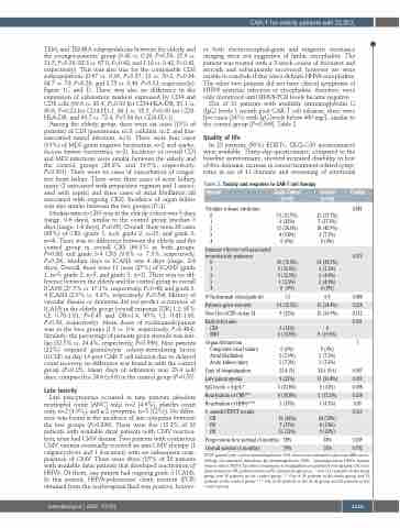
Among the elderly group, there were six cases (15% of patients) of CDI (pneumonia, n=3; cellulitis, n=2, and line- associated tunnel infection, n=1). There were four cases (10%) of MDI (gram negative bacteremia, n=2 and staphy- lococcus hominis bacteremia, n=2). Incidence of overall CDI and MDI infections were similar between the elderly and the control groups (26.8% and 19.5%, respectively, P=0.301). There were no cases of exacerbation of conges- tive heart failure. There were three cases of acute kidney injury (2 associated with preparative regimen and 1 associ- ated with sepsis) and three cases of atrial fibrillation (all associated with ongoing CRS). Incidence of organ failure was also similar between the two groups (P=1).
Median time to CRS was in the elderly cohort was 3 days (range, 0-6 days), similar to the control group (median 3 days [range, 1-6 days], P=0.65). Overall, there were 28 cases (68%) of CRS (grade 1, n=9; grade 2, n=15, and grade 3, n=4). There was no difference between the elderly and the control group in overall CRS (69.3% in both groups, P=0.88) and grade 3-4 CRS (9.8% vs. 7.3%, respectively, P=0.29). Median days to ICANS was 4 days (range, 2-8 days). Overall, there were 11 cases (27%) of ICANS (grade 1, n=5; grade 2, n=5, and grade 3, n=1). There was no dif- ference between the elderly and the control group in overall ICANS (27.5% vs. 17.1%. respectively, P=0.48) and grade 3- 4 ICANS (2.5% vs. 4.9%, respectively, P=0.54). History of vascular disease or dementia did not predict occurrence of ICANS in the elderly group (overall response [OR] 1.2, 95% CI: 0.78-1.81, P=0.45 and OR=1.4, 95% CI: 0.81-1.63, P=0.38, respectively). Mean doses of tocilizumab/patient was in the two groups (1.5 vs. 0.9, respectively, P=0.484). Similarly, the percentage of patients given steroids was sim- ilar (32.5% vs. 24.4%, respectively, P=0.596). Nine patients (22%) required granulocyte colony-stimulating factor (GCSF) on day 14 post CAR-T cell infusion due to delayed count recovery, no difference was found in with the control group (P=0.15). Mean days of admission was 23.4 (±8) days, compared to 24.6 (±9.6) in the control group (P=0.55).
Late toxicity
Late pancytopenia occurred in nine patients (absolute neutrophil count [ANC] only, n=2 [4.9%], platelet count only, n=2 [4.9%], and ≥ 2 cytopenia, n=5 [12%]). No differ- ence was found in the incidence of late cytopenia between the two groups (P=0.399). There were five (15.2% of 32 patients with available data) patients with CMV reactiva- tion, none had CMV disease. Two patients with continuous CMV viremia eventually received an anti-CMV therapy (1 valgancyclovir and 1 foscarnet) with no subsequent reap- pearance of CMV. There were three (15% of 20 patients with available data) patients that developed reactivation of HHV6. Of them, one patient had ongoing grade 3 ICANS. In this patient, HHV6-polymerase chain reaction (PCR) obtained from the cerebrospinal fluid was positive, howev-
er both electroencephalogram and magnetic resonance imaging were not suggestive of limbic encephalitis. The patient was treated with a 3-week course of foscarnet and steroids and subsequently recovered, however we were unable to conclude if this was a definite HHV6 encephalitis. The other two patients did not have clinical symptoms of HHV6 systemic infection or encephalitis, therefore, were only monitored until HHV6-PCR levels became negative.
Out of 21 patients with available immunoglobulin G (IgG) levels 1 month post CAR-T cell infusion, there were five cases (24%) with IgG levels below 400 mg/L, similar to the control group (P=0.398), Table 2.
Quality of life
In 23 patients (56%) EORTC QLQ-C30 questionnaires were available. Thirty-day questionnaire, compared to the baseline questionnaire, showed increased disability in four of five domains, increase in cancer/treatment-related symp- toms in six of 11 domains and worsening of emotional
Table 2. Toxicity and response to CAR-T cell therapy
Domain
Cytokine release syndrome 0
Study Cohort (n=41)
13 (31.7%) 9 (22%) 15 (36.6%) 4 (9.8%)
Control (n=41)
13 (31.7%) 7 (17.1%) 18 (43.9%) 3 (7.3%) 0 (0%)
34 (82.9%) 3 (7.3%) 2 (4.9%) 2 (4.9%) 0 (0%)
0.9
10 (24.4%) 15 (36.9%)
P-value 0.881
0.475
0.484 0.258 0.112 0.301
1
0.547 0.399 0.398 0.234 0.09 0.337
0.209 0.792
1
2
3
4 0(0%)
Immune effector cell-associated neurotoxicity syndrome
0 1 2 3 4
N Tocilizumab (dose/patient) Patients given steroids
Need for GCSF on day 14
29 (72.5%) 5 (12.5%) 5 (12.5%) 1 (2.5%) 0 (0%)
1.5
14 (32.5%) 9 (22%)
Early infections
CDI 5(12%) 0
MDI
Organ dysfunction Congestive heart failure Atrial fibrillation
Acute kidney injury
Days of hospitalization
Late pancytopenia
IgG levels < 4 gr/L*
Reactivation of CMV**
Reactivation of HHV6***
1- month PET/CT results CR
PR PD
6 (14.8%)
0 (0%) 3 (7.3%) 3 (7.3%)
23.4 (8)
9 (22%) 5 (23.8%) 6 (18.8%) 3 (15%)
19 (46%) 7 (17%) 13 (32%)
8 (19.5%)
0 (0%) 3 (7.3%) 3 (7.3%)
24.6 (9.6) 11 (26.8%) 5 (33%) 5 (15.2%) 1 (4.3%)
24 (59%) 8 (19%) 9 (22%)
54% 76%
Progression-freesurvival(6months) 39% Overall survival (6 months) 74%
GCSF: granulocyte colony stimulating factor; CDI: clinical documented infections; MDI: micro- biology documented infections; Ig: immunoglobulin; CMV: cytomegalovirus; HHV6: human herpes virus 6; PET/CT: positron emmission tomography/computerized tomography; CR: com- plete remission; PR: partial remission; PD: disease progression. * Out of 21 patients in the study group and 15 patients in the control group. ** Out of 32 patients in the study group and 33 patients in the control group. *** Out of 20 patients in the study group and 23 patients in the control group.
haematologica | 2022; 107(5)
1115
