
Page 43 - 2022_03-Haematologica-web
P. 43
Genome complexity in CLL: karyotype versus microarrays
AB
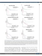
Figure 3. Kaplan-Meier plots for time to first treatment of the genomic risk stratification within each category defined by the alternative technique. (A) Patients classified in each category based on total number of aberrations found by chromosome banding analysis (CBA): non-complex Karyotype (CK) (0-2 abnormalities [abn.]), low/intermediate-CK (3-4 abn.) or high-CK (≥5 abn.) are represented in different plots. Time to first treatment (TTFT) of genomic microarrays (GM) defined groups was assessed. Within non-CK and low/intermediate-CK, cases classified as high-GC (≥5 copy number abnormalities [CNA] by GM) showed a poor outcome. In the high-CK group, those low-GC patients did not display a better evolution while intermediate-GC cases showed an unexpected median TTFT of 22 months. (B) Each plot represents patients classified in each category based on total number of CNA detected by GM: low-GC (0-2 CNA), intermediate-GC (3-4 CNA) or high-GC (≥5 CNA). Within each subgroup, TTFT of CBA defined groups was assessed. Low-GC patients could be stratified in three risk categories when reclassified by CBA, while no significant differences were observed when intermediate-GC and high-GC subsets were reclassified.
reclassification within the low-GC patients allowed the dis- tinction of three risk categories showing similar outcomes to those observed when applied to the global cohort (P<0.001). No significant differences were observed when the intermediate-GC and high-GC categories were reclassi- fied (Figure 3B). It is noteworthy that the ten cases catego- rized in opposite risk groups displayed the poor prognosis
predicted by the technique that classified them in the high- er risk category.
Expectedly, the frequency of TP53 abnormalities (dele- tions and/or mutations) increased together with the com- plexity by both methods. In contrast, intermediate and high risk categories showed a similar increased proportion of unmutated IGHV (U-IGHV) and del(11q) compared
haematologica | 2022; 107(3)
599
