
Page 195 - 2022_03-Haematologica-web
P. 195
Letters to the Editor
p.Gly447Arg, p.Gly506Phe, p.Val516Arg, p.Leu517Pro, p.Thr575Arg, and p.Gly578Ser) were regarded as specif- ic variants associated with isolated thrombocytopenia. Of note, these seven variants spread over a region of approximately 130 amino acids of the ManNac kinase domain (Figure 3).
We analyzed the structure of N-acetylmannosamine kinase in complex with N-acetylmannosamine and ADP (2yhy)12 to assess the potential effect of the mutations on the structure and function of the enzyme (Figure 3). Residues Asp444, Gly447 and Thr575 in the ADP pock- et, and Gly506 and Gly578 close to the substrate domain
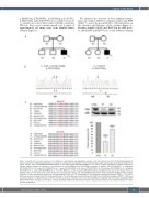
A
B
C
Figure 1. Identification of novel mutations of the GNE gene. (A) Pedigrees and segregation analysis in the two families F1 and F2. (B) Electropherograms of exon 9 showing the c.1546_1547delinsAG and c.1724C>G substitutions in probands P1 and P2, respectively. Sanger sequencing was performed using the fol- lowing primers: 9F/5’-TTCTAGAAATCTTTAAGGTGCTATGG-3’ and 9R/5’-CCACCTGACCATGTTGAAGA-3’. (C) Protein multiple alignments, showing conservation through different species at residues (in red) affected by the p.Val516Arg and p.Thr575Arg mutations. H. sapiens (NP_001121699.1), P. troglodytes (XP_003312121.1), M. mulatta (XP_001082113.2), C. lupus (XP_003431623.1), B. taurus (NP_001178072.2), M. musculus (NP_056643.3), R. norvegicus (NP_446217.1), G. gallus (NP_001026603.2), D. rerio (NP_957177.1), and X. tropicalis (NP_001072728.1). (D) Western blot and of total lysates from lym- phoblast cells of P1 and P2. Total protein lysates were prepared from these cells using M-PERTM Mammalian Protein Extraction Reagent (Thermo Fisher Scientific). Protein quantification shows only a partial expression (39% and 79%, respectively) of GNE expression compared to wild-type (CTRL) (***P<0,002). Actin was used as a loading control for protein quantification. The antibodies were used as follows: anti-GNE (Santa Cruz Biotechnology, sc-376057, 1:500) and anti-β-actin (Santa Cruz Biotechnology, sc-47778, 1:4,000) as primary antibodies, anti-mouse immunoglobulin conjugated with horseradish peroxidase (HRP) (Bethyl, A90-116P, 1:10,000) as a secondary antibody. Statistical analysis was performed using the t-test. Error bars represent the standard deviation of 4 inde- pendent experiments.
D
haematologica | 2022; 107(3)
751
