
Page 192 - 2022_03-Haematologica-web
P. 192
Letters to the Editor
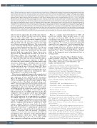
Figure 3. Multiple myeloma tumor growth and dissemination upon immobilization. (A) Right and left hind legs in the same mice were subjected to sciatic den- ervation (DN) and sham operation (control), respectively. Two weeks later, luciferase-transfected mouse 5TGM1 multiple myeloma (MM) cells were simultane- ously inoculated into tibiae in both immobilized (right) and intact (left) hind legs in the same mice. The TAK1 inhibitor LL-Z1640-2 or the PIM inhibitor SMI16a were intraperitoneally injected at 20 mg/kg twice a week. Control groups were given saline as a vehicle. IVIS images taken at 4 weeks. Tumor areas with lumi- nescence shown in green, yellow and red were measured. Px: pixel. Data are expressed as the mean ± standard deviation (SD) (n=5). **P<0.01. (B) 5TGM1 MM cells were inoculated into right tibiae 2 weeks after DN or sham operation. The right tibiae with tumor lesions were harvested at 4 weeks after the MM cell inoculation. Cell lysates were then collected from MM tumor lesions, and protein levels of the indicated factors were analyzed by western blotting analysis. β- actin was used as a loading control. The following reagents were purchased from the indicated manufacturers: antibodies against phosphor-MAP3K7 (Thr187) from Cusabio (Cusabio Biotech, Wuhan, China); and antibodies against TAK1, PIM2, phosho-Smad2, Smad2, and β-actin from Cell Signaling Technology. (C) 5TGM1 MM cells transfected with green fluorescent protein (Gfp) or red fluorescent protein (Rfp) genes were inoculated into tibiae of right hind legs with DN and left sham-operated hind legs, respectively. The GFP-expressing 5TGM1/luc (5TGM1-GFP/Luc) cell line and the RFP-expressing 5TGM1/luc (5TGM1-RFP/Luc) cell line were generated by lentiviral transduction with the pLKO.1-puro-CMV-TurboGFP vector and pLKO.1-puro-CMV-TagRFP vector (Sigma-Aldrich, MO, USA), respectively. Four weeks later, IVIS images were taken. Blood was drawn from retro-orbital plexus, and tumors detected in the IVIS images were resected with surrounding tissues in the mice. Tumors emitting green or red fluorescence were visualized in resected samples with a fluorescence microscope (OLYMPUS SZX16). (D) Circulating 5TGM1-GFP and 5TGM1-RFP cells were analyzed in the blood samples from the mice at 4 weeks by flow cytometery.
tumorous lesions appeared in mice with sciatic denerva- tion over time at around 4 weeks or later at sites distant from the tibiae where MM cells were inoculated. In order to better analyze the metastatic expansion of MM cells inoculated into the tibiae, we transfected 5TGM1 MM cells with either the green fluorescent protein (Gfp) or red fluorescent protein (Rfp) gene. The in vitro prolif- eration of the Gfp- and Rfp-transfected MM cells was the identical (Online Supplementary Figure S2C). The Gfp- and Rfp-transfected cells were inoculated into tibiae of the right hind legs with sciatic denervation and left sham- operated hind legs, respectively. Tumor lesions were detected at sites distant from the tibiae at 4 weeks in IVIS images (Figure 3C). Interestingly, all tumorous lesions metastasized outside of the tibiae were found to be composed of the MM cells labeled with GFP, indicat- ing preferential extraosseous expansion of MM cells inoculated into tibiae in hind legs paralyzed with sciatic denervation. Furthermore, substantial numbers of GFP- positive cells but not RFP-positive cells were detected in sera drawn from the mice at 4 weeks (Figure 3D). These results demonstrate the acceleration of MM tumor growth with egression from the bone marrow into circu- lation and thereby extraosseous dissemination under immobilization or mechanical unloading.
There were no significant changes in the serum levels of sclerostin (Online Supplementary Figure S3B) as well as Sost gene expression in mice with the sciatic denervation (Figure 1F). Robling et al. investigated the mechanoregula- tion of Sost mRNA and sclerostin under enhanced (ulnar loading) and reduced (hindlimb unloading) loading condi- tions.10 Sost transcripts and sclerostin protein levels were significantly reduced at 24 hours in the ulna fixed to the loading platens and actuator after 360 cycles of mechani- cal loading per day. In contrast, mice subjected to tail sus- pension (hindlimb unloading) for 3 days exhibited a sig- nificant increase in Sost mRNA expression in the tibia compared to those in ground control mice. Intriguingly, this upregulation subsided to be non-significant after 7 days of tail suspension. In our experiments, we analyzed Sost mRNA expression at 14 days after the immobiliza- tion, and found that Sost mRNA was not increased signif- icantly. Sost mRNA induction by mechanical unloading may be temporal and should be studied in a time- sequence manner. However, serum levels of sclerostin have been demonstrated to be increased in MM patients with active bone lesions11,12 and positively correlate with lumbar spinal bone mineral density in postmenopausal women.13 Consistent with the patients’ observation, serum levels of sclerostin were increased after MM cell inoculation in mice, and more in mice with mechanical unloading than in control mice (Online Supplementary Figure S3B), which may be due to the acceleration of MM tumor expansion resulting from mechanical unloading.
Bone is a unique microenvironment for MM cell growth and survival, which provides niches to foster clonogenic and dormant MM cells. The present study demonstrates that hind leg immobilization or mechani- cal unloading aggravates bone destruction and MM tumor expansion. In contrast, mechanical loading with repeated forced compression 14 and low intensity vibra- tion 15 has been reported to suppress osteolysis and the growth of MM cells in bone. In order to keep bone mass in MM, repeated mechanical loading appears to play an important role. These observations warrant further study on the therapeutic merit of mechanical stress or loading in MM.
Kotaro Tanimoto,1 Masahiro Hiasa,1 Hirofumi Tenshin,1 Jumpei Teramachi,2 Asuka Oda,3 Takeshi Harada,3 Yoshiki Higa,1 Kimiko Sogabe,3 Masahiro Oura,3 Ryohei Sumitani,3 Tomoyo Hara,3 Itsuro Endo,4 Toshio Matsumoto,5 Eiji Tanaka,1 and Masahiro Abe3
1Department of Orthodontics and Dentofacial Orthopedics, Tokushima University Graduate School of Biomedical Sciences, Tokushima; 2Department of Oral Function and Anatomy, Okayama University Graduate School of Medicine, Dentistry and Pharmaceutical Science, Okayama; 3Department of Hematology, Endocrinology and Metabolism, Tokushima University Graduate School of Biomedical Sciences, Tokushima; 4Department of Bioregulatory Sciences, Tokushima University Graduate School of Medical Sciences, Tokushima and 5Fujii Memorial Institute of Medical Sciences, Tokushima University, Tokushima, Japan
Correspondence:
MASAHIRO ABE - masabe@tokushima-u.ac.jp MASAHIRO HIASA - mhiasa@tokushima-u.ac.jp doi:10.3324/haematol.2021.278295
Received: January 12, 2021
Accepted: November 11, 2021.
Pre-published: November 18, 2021.
Disclosures: MA received research funding from Chugai Pharmaceutical, Sanofi K.K., Pfizer Seiyaku K.K., Kyowa Hakko Kirin, MSD K.K., Astellas Pharma, Takeda Pharmaceutical, Teijin Pharma and Ono Pharmaceutical, and honoraria from Daiichi Sankyo Company. All other authors declare no competing financial interests.
Contributions:KT, MH and MA designed the research and con- ceived the project; KT, MH, HT, JT, AO, TH, YH, KS, MO and RS conducted animal experiments; TH, IE, TM, ET evaluated muscle and bone specimens; KT, MH, KS, MO and RS conducted in vitro cultures; KT, MH, JT, HT, AO, TH and YH performed immunoblotting and immunohistochemical analyses; KT, MH and AO performed flow cytometric analysis; and KT, MH, JT and HT performed transfection and PCR; KT, MH, IE, TM, ET and MA analyzed the data; KT, MH and MA wrote the manuscript.
748
haematologica | 2022; 107(3)
