
Page 173 - 2022_03-Haematologica-web
P. 173
ZBP1 regulates myeloma cell proliferation via IRF3/IRF4
AB
CDF
E
GH
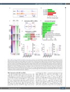
Figure 6. IRF3 co-operates with IRF4 and regulates cell cycle genes in myeloma cells. (A, B) IGV browser snapshots of IRF3 and IRF4 co-binding at the promoter and super-enhancer of IRF4 as assessed by chromatin immunoprecipitation (ChIP)-sequencing (A) and log2 fold-change of IRF4 expression (Padj <0.05) assessed by RNA-sequencing after depletion of indicated mRNA/protein in relation to the scrambled (scr)control in MM.1S cells (B). (C) Heatmaps of IRF3 and IRF4 genome-wide binding, and common binding regions of IRF3 and IRF4 (intersection) as their binding regions are intersected by Bedtools Intersect.50 Numbers of binding regions are shown in brackets. (D) Venn diagram showing the numbers of genomic regions of IRF3 and IRF4 co-binding with gene regulatory potential as assessed by inte- gration of the whole transcriptome of IRF3-depleted MM.1S cells with IRF3- or IRF4-cistrome alone or IRF3 and IRF4 co-binding regions (intersection) in MM.1S cells. Here IRF4 genome-wide binding regions with very low scores were omitted and only the top 50% binding regions with highest scores were used. (E) Numbers of genes predicted to be directly co-regulated (repressed or activated) by IRF3 and IRF4 binding. The co-binding (intersection) regions were integrated with the IRF3-depleted transcriptome by shRNA1 and shRNA2 in MM.1S cells. (F) Enrichr pathway enrichment analysis for common genes predicted to be activated by IRF3-IRF4 co-binding. (G) Primary ChIP quantitative polymerase chain reaction (qPCR) against IRF3 (left) followed by re-ChIP-qPCR against IRF4 (right). The position of amplicons is shown as horizontal colored lines in Figure 6A. (H) Primary ChIP-qPCR against IRF4 (left) followed by re-ChIP-qPCR against IRF3 (right). The position of amplicons is shown as horizontal colored lines in Figure 6A.
ZBP1 interaction with IRF3 and TBK1
We next investigated the downstream processes that might link constitutive ZBP1 expression in myeloma cells with regulation of cell cycle. Unlike in non-malignant cells in which IRF3 phosphorylation/activation requires activa- tion of sensors such as cGAS-STING,14,16 we found that IRF3 was constitutively phosphorylated (pIRF3) in myelo- ma cell lines (Figure 4A). In line with previous reports,39 pIRF3 was also detected in other non-myeloma cancer cells (Figure 4A). Importantly, pIRF3 was also detected in primary BM myeloma CD138+ PC (Figure 4B) and thus
establishing that IRF3 is constitutively phosphorylated in MM. Although the functional relationship of the ZBP1-IRF3 interaction in the context of cellular innate immune responses is a matter of debate, the physical interaction of ZBP1-IRF3 was previously demonstrated by their ectopic expression.17,40,40 Using protein co-immuno- precipitation assays we found that endogenous ZBP1 interacts with endogenous IRF3 (Figure 4C, D) and TBK1 (Figure 4E) in MM.1S cells. By co-transfection of IRF3 cDNA with a full length or C-terminus deleted mutant of ZBP1 (Online Supplementary Figure S5A), we found that the
haematologica | 2022; 107(3)
729
