
Page 79 - 2022_02-Haematologica-web
P. 79
RXRA active mutation
RXRA WT displayed increased leukocytosis, an increase in neutrophils, and a decrease in lymphocytes compared to mice transplanted with RXRA DT448/9PP-transduced cells (Figure 4B-D). Compared with the pre-engraftment popula- tion (RXRA WT, 55% mCherry+; RXRA DT448/9PP, 62% mCherry+), at 4 weeks there was reduced engraftment in RXRA DT448/9PP cells relative to RXRA WT cells: RXRA WT average of 7.6% mCherry+ cells (range, 0.94%-16.4%) versus RXRA DT448/9PP 0.25% mCherry+ cells (range,
0.095%-0.36%) (Figure 4E). When we assessed moribund mice transplanted with cells transduced with RXRA WT, we observed further reduction in the absolute proportion of mCherry+ cells in the bone marrow rather than an expan- sion: RXRA WT, average 1% (range, 1.21%-1.46%); RXRA DT448/9PP, average 0.741 (range, 0.022%-1.46%) (Figure 4G). Survival was shorter in mice transplanted with RXRA WT-transduced cells than in those transplanted with RXRA DT448/9PP-transduced cells (Figure 4H). At the time of sac-
ABC
D
E
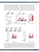
Figure 2. Constitutive activity of RXRA DT448/9PP. (A) KMT2A-MLLT3 WT leukemia cells derived from UAS-GFP bone marrow cells were transduced with MSCV-Gal4- RXRA DT448/9PP-IRES-mCherry or MSCV-Gal4-RXRA-IRES-mCherry, treated with increasing concentrations of bexarotene, and the ratio of GFP+ cells (responding) to total mCherry+ cells (capable of responding) was determined. Data from biological duplicates are shown. (B, C) 293T cells were transfected with the indicated plas- mids, treated with bexarotene, HX531, or UVI3003 (all 1 mM), and luciferase was measured after 40 h. Data from biological triplicates are shown. PPRE: peroxisome proliferator response element. ApoA1: DR1 element from the ApoA1 promoter. RARE: retinoic acid receptor response element from the RARB promoter. The ∆AF2 deletion acts as a negative control. (D, E) Mammalian two-hybrid. 293T cells were transfected with the indicated plasmids, treated with bexarotene and GFP was measured by flow cytometry after 40 h. Data from biological triplicates are shown. GFP-N1: a positive control CMV-GFP expression vector. GFP-N0: a negative control derived from GFP-N1 after the deletion of the CMV promoter. Statistical significance was evaluated using analysis of variance with the Bonferroni comparison to con- trol, *P<0.05, **P<0.01, ***P<0.001.
haematologica | 2022; 107(2)
421
