
Page 217 - 2022_02-Haematologica-web
P. 217
Letters to the Editor
expression of MK sterile a motif (SAM) and histidine- aspartate (HD) domain containing deoxynucleoside triphosphate triphophohydrolase 1 (SAMHD1) and (ii) SAMHD1 expression inhibits cultured human MK pro- platelet formation (PPF) and promotes apoptosis. This is the first identification of SAMHD1 in human MK and report of a dNTP hydrolase regulating platelet produc- tion.
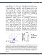
In order to pursue studies on the effects of inflamma- tion on megakaryocytopoiesis, we used CD34+ hematopoietic stem cells derived from human umbilical vein cord blood. IFNa significantly decreased day 13 PPF and platelet-like particles (Figure 1A to C), but did not affect the percentages of MK or polyploidy (Figure 1D to E). Importantly, we also showed that exogenous IFNa induces thrombocytopenia in wild-type mice (Figure 1F), consistent with studies in immunodeficient mice.4 In order to begin to understand how IFNa regulates late- stage megakaryopoiesis and platelet production, we used an unbiased, transcriptome-wide approach and per- formed RNA sequencing (RNA-seq) on CD61-purified, day 13 cultured MK stimulated with IFNa. Our analyses identified 201 transcripts that were differentially expressed at a nominal significance threshold (P<0.05). Adjusting for multiple comparisons and setting a false discovery rate (FDR) threshold of <0.05, we found that 66 of the 201 transcripts were upregulated by IFNa (Online Supplementary Table S1). Increased mRNA expression in response to IFNa was validated by real- time polymerase chain reaction (PCR) analysis for all five genes tested (SAMHD1, PHF11, ISG20, IFITM3 and TAP2) (Online Supplementary Figure S1). Gene ontology analysis indicated that the differentially expressed genes were associated with the type 1 interferon signaling pathway, defense response to virus, and negative regula- tion of viral genome replication. Subsequent studies focused on SAMHD1, whose abundance increased more than 16-fold with IFNa induction (FDR=2.0x10⁻¹⁸)
(Figure 2A). Figure 2B and C shows that IFNa treatment of cultured MK greatly increased the abundance of SAMHD1 mRNA and protein (n=3 independent biologi- cal replicates). SAMHD1 is a hydrolase, the activated form of which degrades the intracellular pool of deoxynucleoside triphosphates (dNTPase) into deoxynu- cleosides and inorganic triphosphates, and is known to restrict viral replication of the human immunodeficiency virus type-1.8 In addition to viral restriction, SAMHD1 is required for cellular functions including replication fork progression, cell proliferation, apoptosis and DNA dam- age repair.9 IFNa stimulation induces SAMHD1 expres- sion in human monocytes,10 astrocytes, microglia,11 HEK293T and HeLa cells,12 but there are no prior reports of SAMHD1 expression and/or function in MK or platelets.
Platelet RNA And eXpression 1 (PRAX1) data13 demonstrated that SAMHD1 transcript levels are nega- tively associated with platelet count in healthy human subjects (Figure 3A), suggesting a possible inhibitory role of SAMHD1 in platelet production. Since SAMHD1 modulates the intracellular levels of dNTP, we hypothe- sized that an increase in the abundance SAMHD1 upon IFNa stimulation leads to decreased MK proliferation, maturation and DNA synthesis (MK polyploidy). However, deletion of SAMHD1 by CRISPR/Cas9 gene editing in cultures promoting unilineage MK differentia- tion (Figure 3B) did not affect MK maturation (Online Supplementary Figure 2A and B) or ploidy (Online Supplementary Figure 2C). This suggests SAMHD1 effects thrombopoiesis rather than megakaryocytopoiesis. Similar to Figure 1, IFNa stimulation caused a significant decrease in MK PPF MK without CRISPR modification (Figure 3C, first 2 bars). The effect of IFNa on PPF was abolished when SAMHD1 was deleted (Figure 3C, sec- ond 2 bars). Lastly, IFNa is well-established as pro-apop- totic.2 MK must restrain apoptosis to survive and progress safely through PPF and platelet shedding.14,15
ABC
Figure 2. Interferon α upregulates. dNTP hydrolase enzyme SAMHD1 in cultured megakaryocytes. CD34+ hematopoietic stem cells and progenitor cells were isolated by immunomagnetic separation from human umbilical cord blood. Cells were cultured in megakaryocyte (MK) promoting conditions by supplementation with thrombopoietin for 13 days. 1,000 units per milliliter (U/mL) of interferon α (INFα) and phosphate-buffered saline (PBS) (negative control) was added in 3 independent cultures at day 13 for 8 hours. (Dose and time course studies were performed in Meg-01 to select optimal concentrations and times, data not shown). CD61+ MK were separated by immunomagnetic beads and total RNA was obtained by mirVana kit. RNA samples with RIN score of >8 were used for RNA sequencing. The library preparation was done using TruSeq standard total RNA sample preparation kit with RNA depletion (Illumina Inc.). RNA sequensing was performed using NextSeq500 instrument with read length of 50 basepair single-end read. On average 68.9 million reads were obtained from each sample and genome mapping was on average 94.5% for all samples. (A) Volcano plot shows the relationship between the P-values and the fold change in normalized expression for IFNa or PBS treated cultured human MK. Differentially expressed genes (false discovery rate [FDR] <0.05) on IFNa treatment was plotted. SAMHD1 is shown in red. (B) Real-time polymerase chain reaction analysis validates the upregulation of SAMHD1 in IFNa-treated CD61+ MK compared to PBS control. Actin was used as housekeeping gene. Log2 fold change of SAMHD1 mRNA levels is plotted for IFNa vs. PBS control (n=3). Statistical significance was determined by one sample t-test. (C) Representative immunoblot shows increase in SAMHD1 protein in IFNa-treated MK compared to PBS control. Actin is used as loading control.
haematologica | 2022; 107(2)
559
