
Page 210 - 2022_02-Haematologica-web
P. 210
Letters to the Editor
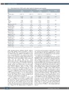
Table 1. Clinical characteristics of HTLV-2 positive subjects analyzed in immunogene panel sequencing.
Mutation Number
Total 0 1-2 >3 P-value
n=28(%) n=10(36) n=8(29) n=10(36)
Age
Median(range) 53(43-70) 52(46-70) 54(43-70) 60(49-68) 0.04†
Sex Female
Male
Race Black
Hispanic White
White blood cells (109/L) Median (range)
Hemoglobin (g/dL) Median (range)
Hematocrit (%) Median (range)
Platelets (109/L) Median (range)
Lymphocytes (109/L) Median (range)
Eosinophils (109/L) Median (range)
Neutrophils (109/L) Median (range)
Monocytes (109/L) Median (range)
Basophils (109/L) Median (range)
19 (68) 9 (32)
9 (32) 5 (18) 14 (50)
6.2 (3.5-10)
13.9 (4.8-17.4)
40.8 (14-50.5)
231 (101-435)
2.378 (0.984-4.122)
0.130 (0.028-0.319)
3.207 (1.404-6.232)
0.410 (0.042-1.140)
0.0 (0.0-0.2)
7 (70) 3 (30)
1 (10) 2 (20) 7 (70)
6.2 (3.5-8.2)
13.4 (4.8-16.7)
40.3 (14-49.2)
222 (144-435)
1.906 (0.984-3.854)
0.117 (0.028-0.246)
3.418 (1.582-6.232)
0.378 (0.042-0.711)
0.0 (0.0-0.2)
6 (75) 2 (25)
4 (50) 1 (12) 3 (38)
6.6 (6.0-9.1)
13.6 (12.5-17.4)
39.9 (38.7-50.5)
239 (200-340)
2.907 (1.365-4.122)
0.185 (0.130-0.319)
3.393 (2.500-4.940)
0.434 (0.195-0.919)
0.0 (0.0-0.1)
6 (60) 4 (40)
4 (40) 2 (20) 4 (40)
5.8 (3.9-10)
14.2 (12.8-16.2)
42.7 (38.1-48.9)
240 (101-350)
2.378 (1.566-3.610)
0.078 (0.057-0.200)
2.944 (1.404-5.700)
0.480 (0.235-1.140)
0.0 (0.0-0.2)
0.63^
0.41^
0.85† 0.27† 0.15† 0.80† 0.24† 0.21† 0.51† 0.24† 0.99†
The data are at sample collection. P-values are calculated by Cuzick’s trend test (†), or Kruskal-Wallis trend test (^).
STAT signaling pathway (NFKBIA, PIK3R5, MAPK14, EP300, MPL, IFNAR1, IL6ST and IL20RA) according to Uniprot identifier. Three genes had more than one vari- ant: VWF (3 mutations), SMAD7 and MXRA5 (2 muta- tions each) (median VAF: 7%, 2% and 4%, respectively). Subject 13 who harbored a STAT3 Y640F mutation also had mutations in KMT2D, NFKBIA, PIK3R5, CTCF, and VWF. In this subject with multiple somatic mutations, STAT3 had the highest VAF (16.2%, Online Supplementary Table S1), suggesting that other variants may be subclonal. Subject 12 with a STAT3 N647I muta- tion also harbored variants in INPP5D, FSCN1 and PLA2R1, CD248, and P4HTM. Subject 11 with STAT3 Y657_K658insY harbored variants in MTA1, NCOR2, BCL6, ADCY8, RPS6KA3. Subject 27 with a STAT3 D661Y mutation had no additional variants. The overall number of mutations was higher in HTLV-2 positive blood donors harboring STAT3 mutations (median =6) compared to HTLV-2 positive blood donors without STAT3 mutations (median =1; P=0.061; Mann–Whitney U test), although the difference was not statistically sig- nificant. The complete list of variants and VAF can be found in the Online Supplementary Table S1.
Somatic mutations can accumulate in tissues with aging.14 Accordingly, the total number of coding variants was associated with older age among HTLV-2 positive blood donors (no variants, median age 51 years; 1-2 vari- ants, 53 years; 3 or more variants, 59 years; P=0.04) (Table 1; Figure 2B). The most frequent single nucleotide transi- tion was C>T involved in age-associated mutational signa- ture 1 (Figure 2C). The high prevalence of signature 1,
revealed by mutational signature analysis (Figure 2D), fur- ther supported age-dependent accumulation of somatic variants in CD8+ T cells among HTLV-2 positive subjects. No association between peripheral blood counts and num- ber of variants was observed (Table 1).
In summary, our results highlight the presence of STAT3 mutations in CD8+ T cells of healthy blood donors harbor- ing HTLV-2 without clinical history of lymphoproliferative disease. HTLV-2 positive subjects with STAT3 mutations showed variable clonal expansion of CD8+ T cells, sug- gesting that HTLV-2 infection may promote lymphoprolif- eration and STAT3 mutagenesis outside the clinical context of T-LGLL. We identified additional mutations in CD8+ T cells of HTLV-2 positive subjects in genes involved in JAK- STAT signaling, immune regulation and lymphoprolifera- tion. In addition to T-LGLL, somatic STAT3 mutations have been detected in LGLL associated diseases such as aplastic anemia, hypoplastic myelodysplastic syndrome and Felty’s syndrome, and in some patients with multiple sclerosis and rheumatoid arthritis.15 Although CD8+ T cell expansions can also be detected in other diseases such as in rheumatoid arthritis, somatic STAT3 mutations are not common in these conditions.8 STAT3 mutations and a his- tory of HTLV-2 infection may highlight a subset of blood donors who are at risk of subsequent diagnosis of lympho- proliferative diseases. However, no clinical follow-up information is available from our study participants, and future studies are needed to elucidate whether HTLV-2 positive subjects carrying STAT3 and other mutations are at increased risk of T-LGLL or other lymphoproliferative diseases.
552
haematologica | 2022; 107(2)
