
Page 140 - 2022_02-Haematologica-web
P. 140
L. Traeger et al.
siNDFIP1 successfully depleted NDFIP1 mRNA in both the absence (Figure 3B) and the presence (Online Supplementary Figure S3C) of exogenous BMP6. siRNA directed against NDFIP1 had no effect on the ability of BMP6 to induce hep- cidin expression, demonstrating that the BMP signaling pathway was intact (Figure 3C). To confirm that depletion of NDFIP1 blocks degradation of FPN-GFP, NDFIP1 deplet-
ed cells were treated with exogenous hepcidin (4 ng/mL). Compared to cells that were transfected with siControl, depletion of NDFIP1 inhibited hepcidin-mediated degrada- tion of the FPN-GFP fusion protein (Figure 3D; Online Supplementary Figure S2C).
To investigate the possibility that NDFIP1 interacts with ferroportin, HepG2 cells were incubated in the presence or
A
B
C
DE
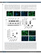
Figure 2. UBA6 is required for hepcidin-mediated degradation of the ferroportin-green fluorescent protein fusion protein. (A) Cells were transfected with siControl, siUBA1 or siUBA6, and treated with doxycycline (Dox) or Dox followed by BMP6 (10 ng/mL) for 18 hours (h), as indicated. Dox-induced expression of ferroportin-green fluorescent protein (FPN-GFP); depletion of UBA6, but not UBA1, prevented BMP6-mediated FPN degradation. (B) The levels of FPN-GFP and phosphorylated SMAD 1/5/8 in siControl-, siUBA1- and siUBA6-transfected HepG2-FPN-GFP cells in the presence (+) or absence (-) of BMP6 (10 ng/mL for 18 h) are shown. In the absence of Dox, control cells did not express the FPN-GFP fusion protein. Treatment with BMP6 increased the level of pSMAD 1/5/8. UBA6 depletion prevented BMP6-medi- ated degradation of FPN-GFP. GAPDH was used as a loading control. The immunoblot is representative of 3 separate experiments. (C) UBA1 and UBA6 were success- fully depleted using the appropriate small interfering RNA (siRNA), as determined by quantitative polymerase chain reaction (qPCR) (mRNA expression relative to con- trol; *P<0.05; **P<0.01; Mann-Whitney-U test). (D) Treatment with BMP6 (10 ng/mL for 18 h) induced the expression of hepcidin in siControl-transfected cells. Depletion of SMAD4, but not depletion of UBA6, blunted the BMP6-mediated induction of hepcidin in HepG2-FPN-GFP cells (mRNA expression relative to control; **P<0.01; Kruskal-Wallis test). (E) Images of cells transfected with siControl, siUBA1 and siUBA6 are shown. Cells were treated with Dox or Dox followed by hepcidin (4 ng/mL for 18 h), as indicated. Treatment with Dox induced expression of FPN-GFP in siControl treated cells, while incubation with hepcidin caused the degradation of the fusion protein. Depletion of UBA6, but not UBA1, prevented hepcidin-mediated degradation of the FPN-GFP fusion protein. The location of nuclei in (A) and (E) are indicated by staining with DAPI (blue). White bar indicates 100 μm.
482
haematologica | 2022; 107(2)
