
Page 139 - 2022_02-Haematologica-web
P. 139
Regulation of ferroportin degradation
A
E
B
D
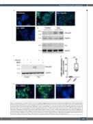
Figure 1. Characterization of the HepG2-FPN-GFP cell line. (A) Images of untreated HepG2 cells, and cells treated with doxycycline (Dox; 2 μg/mL) alone, with Dox followed by hepcidin (50 ng/mL) for 90 minutes (min), with Dox followed by BMP6 (10 ng/mL) for 18 hours (h) are shown. (B) Treatment with Dox induced the expres- sion of the ferroportin-green fluorescent protein (FPN-GFP) fusion protein. Dox-treated cells had reduced levels of intracellular ferritin light-chain (FTL), consistent with increased iron export in cells expressing FPN-GFP. GAPDH was used as a loading control. (C) In the absence of Dox, the fusion FPN-GFP protein was not detected by immunoblot (lanes 1 to 3). In the presence of Dox, FPN-GFP was expressed (lane 4). Treatment with hepcidin (50 ng/mL; lane 5), or BMP6 (10 ng/mL; lane 6), for 18 h caused degradation of the FPN-GFP fusion protein. (D) BMP6 stimulation (10 ng/mL) for 18 h induced hepcidin mRNA expression in HepG2-FPN-GFP cells, as determined by quantitative polymerase chain reaction (qPCR) (mRNA expression relative to control; **P<0.01; Mann-Whitney-U test). (E) Images of siSMAD4 trans- fected cells treated with Dox (left panel), siSMAD4 transfected cells treated with Dox followed by BMP6 (10 ng/mL; middle panel) and siControl transfected cells treat- ed with Dox followed by BMP6 (right panel) are shown. Small interfering RNA (siRNA)-mediated inhibition of SMAD4 prevented BMP6-mediated degradation of the FPN-GFP fusion protein. The location of nuclei in (A) and (E) are indicated by staining with DAPI (blue). White bar indicates 100 μm.
haematologica | 2022; 107(2)
481
C
