
Page 111 - 2022_02-Haematologica-web
P. 111
Gene therapy of DBA
Discussion
Currently, hematopoietic stem cell transplantation is the sole curative option for DBA patients, but suitable donors are often unavailable and there can be serious immunological complications.3,33 Gene therapy using gene-corrected hematopoietic stem cells has been shown to be a promising therapeutic strategy for genetic blood disorders in recent years.19,34,35 Our previous proof-of-con- cept studies also demonstrated the feasibility of applying gene therapy to cure DBA.12-16 In the present study, a clin- ically applicable lentiviral vector was used to investigate the efficacy and safety for treating anemia and lethal BM failure in Rps19-deficient mice and for ameliorating the impaired erythroid differentiation in human primary RPS19-deficient CD34+ cord blood cells. The next step towards clinical gene therapy will be to perform toxicol- ogy and biodistribution analyses, and thereafter proceed with submitting an application to regulatory authorities in order to initiate a phase I/II clinical trial with 6-12 patients focusing on safety.
For successful development of clinical gene therapy, vec- tor efficacy, generating long-term therapeutic effects, is crucial. To examine the therapeutic effects of the vector with lower copies per cell, we decreased the MOI to 5-10 in the present study and demonstrated that the EFS-RPS19 vector has robust therapeutic effects with no evidence of clonal expansion associated with vector integration near cancer-associated genes, as we showed in our previous study.12 It has already been shown that ribosomal proteins are produced in excess of the needs of the ribosome assembly, and that the excess protein is subjected to pro- teasomal degradation.22,29,30 Similarly, in the present study, our transduced Rps19-deficient cells had physiological lev- els of expression of RPS19. Hence, it is unlikely that the ectopic expression of RPS19 would promote uncontrolled
growth. Since single-copy insertion of the therapeutic gene in the target cells is suggested to avoid the risk of genotoxicity in clinical gene therapy manipulation,19 we plan to examine the therapeutic effects using transduced cells with a lower MOI (e.g. MOI=1) in future studies. Our results also demonstrated that the vector could rescue the impaired erythroid differentiation of RPS19-deficient cord blood cells by increasing red blood cell production. Overall, we showed that the EFS-RPS19 vector could res- cue the anemia and BM failure of RPS19-deficient DBA.
Apart from efficacy, vector safety is the other essential factor to assess when applying gene therapy. The risk of insertional mutagenesis is a concern for future applications of gene therapy in the clinic. To prevent this risk, we uti- lized a third-generation SIN lentiviral vector that lacks potent enhancers in the long terminal repeat regions, since such vectors were shown to exhibit a safer integration profile in previous clinical trials19,34,36 and also in our previ- ous animal studies.12,15 By using the state-of-the-art INSPI- IRED workflow, which can provide better quantification of clonal abundance compared to a linear amplification- mediated PCR approach, we found that gene-corrected BM cells in both models exhibited a low risk of mutagen- esis with no evidence of clonal expansion associated with vector integration near cancer-associated genes. In our study, no hematologic abnormalities were observed due to enforced expression of RPS19. The results collectively demonstrate the safety of the EFS-RPS19 vector for clinical gene therapy development. The bioinformatic pipeline of the INSPIIRED workflow is a more automated approach, making it well suited for monitoring patients in gene ther- apy trials in the future.
Increased MCV, due to macrocytic anemia, is a classic clinical observation in patients with DBA. It is, at least in part, caused by the stabilization of p53 and activation of p53 targets (e.g., p21, Bax), which are responsible for cell
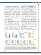
ABCD
EFGHI
Figure 6. EFS-RPS19 vector-treated Rps19-deficient cells show a competitive advantage in contributing to long-term hematopoiesis in vivo. (A, B) Vector copy num- ber in peripheral blood (A) and bone marrow (B). (C, D) Donor reconstitution in peripheral blood (C) and bone marrow (D). (E–I) The percentage of transduced cells in hematopoietic stem cells (E), megakaryocyte progenitors (F), pre-granulocyte-macrophage and granulocyte-macrophage progenitors (G), pre-megakaryocyte-ery- throid (H), and pre-colony-forming unit erythroid and colony-forming unit erythroid (I) (n=14-16, error bars represent the standard deviation, black asterisks indicate the statistical significance of the comparison of recipient-derived cells between the mock and EFS-RPS19 groups, orange asterisks indicate the statistical signifi- cance of the comparison of donor-derived cells between the mock and EFS-RPS19 groups. *P<0.05, **P<0.01, ***P<0.005, ****P<0.001 by one-way analysis of variance). VCN: vector copy number; PB: peripheral blood; BM: bone marrow; HSC: hematopoietic stem cells; MkP: megakaryocyte progenitors; pre-GM/GMP: pre- granulocyte macrophage and granulocyte macrophage progenitors; preMegE: pre-megakaryocyte-erythroid; preCFU-E/CFU-E: pre-colony-forming unit –erythroid (CFU- E)/CFU-E.
haematologica | 2022; 107(2)
453
