
Page 289 - 2022_01-Haematologica-web
P. 289
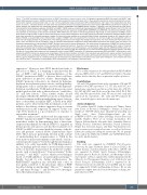
RINF maintenance of SMAD7 sustains human erythropoiesis
Figure 7. The RINF knockdown-induced phenotype is SMAD7-dependent in human primary cells. (A) Histogram representing RINF (left panel) and SMAD7 (right panel) mRNA expression levels in shRNA-transduced CD34+ cells isolated from three cord blood units. In these specific experiments, lentiviral transduction was per- formed after magnetic sorting of CD34+ cells (i.e., day 0 of the amplification step) and cells were GFP-sorted between 2 and 5 days of the amplification step. Data shown are mean ± standard deviation for each donor (the quantitative reverse transcriptase polymerase chain reaction [qRT-PCR] was performed in triplicate). To assess whether the mRNA level was statistically different between shRNA-RINF and shRNA-Scramble samples, a Student t-test was performed for each donor. Considering the three biological replicates, both RINF and SMAD7 mRNA levels were statistically downregulated by shRNA-RINF (paired Student t-test, P<0.001). (B) This histogram represents the level of SMAD7 mRNA obtained with the two sequences targeting RINF (shRINF#3 and shRINF#4) and detected by qRT-PCR. Values are expressed as percentages of the shControl condition. RINF mRNA levels are shown in Online Supplementary Figure S3B. (C) Since detection of SMAD7 protein level requires a large quantity of cells for western blot analysis and the active endogenous level of this protein can be below the detection threshold, SMAD7 protein levels were estimated by immunofluorescence. Images of stained cells were acquired on a wide-field Nikon Eclipse microscope through a ×20 objective, with an Eclipse TE2000, a cascade CDD camera (Photometrics). Images of SMAD7 had to be acquired with a binning of one, and two images were averaged for each con- dition. For cell imaging analysis the quantification of the labeling, performed on two images after background subtraction, the mean fluorescent intensity was meas- ured in 100–120 cells for the two conditions, using Fiji software. Representative images from three independent experiments are shown. Scale bar=50mm. (D) Schematic representation of the pInducer21/SMAD7 lentiviral vector used for rescue experiments. This vector drives the expression of SMAD7/HA under a tetracy- cline response element (TRE) promoter that is activated after doxycycline treatment. To perform rescue experiments, cord blood-derived CD34+ cells (n=3 donors) were first transduced with the pInducer21/SMAD7 lentiviral vector, sorted for a low GFP expression, and transduced again with the pTRIPDU3/shRNA vectors tar- geting RINF or control. After a second step of GFP sorting (this time based on high GFP expression to sort cells transduced with both lentiviral vectors), cells were cultured for 2 more days before adding (or not) doxycycline, which induced SMAD7 expression. (E) Histograms representing RINF and SMAD7 mRNA levels in cells, detected by qRT-PCR. Values are expressed as percentages of the shControl condition. A Student t-test (unpaired) was performed to assess whether the observed relative difference was statistically significant. (F) RINF and SMAD7 were detected by western-blot analysis after doxycycline induction (here after 3 days of treatment with 0.2 μg/mL). ACTB was used as a loading control. (G) Cell maturation was determined by a benzidine assay (upper panel histogram). (H) Cumulative red blood cell number at day 17 of exposure to erythropoietin (EPO) is presented in the histogram in the lower panel.
expression.33 However, since RINF knockdown leads to global loss of 5hmC, it is tempting to speculate that the loss of RINF could lead to hypermethylation of the SMAD7 promoter in HSPC, a process that could have gone unnoticed in previous studies. Interestingly, the SMAD7 promoter is known to be silenced by hyperme- thylation in non-hematopoietic tissues, and hypomethy- lating agents such as azacytidine can revert this hyperme- thylation, and alleviates TGFb-induced diseases in several pathological models such as atherosclerosis,61 renal fibro- sis62 and liver fibrosis.63 Thus, further studies should investigate whether the SMAD7 promoter is demethylat- ed after treatment with hypomethylating agents (azacyti- dine or decitabine) in human HSPC isolated from MDS patients, and to what extend this mechanism could con- tribute to the efficacy of these epigenetic drugs to sustain erythropoiesis in the long-term. In support of this con- cept, TET2 mutation is a good indicator of response to azacytidine treatment.64
Previous studies have underscored the importance of SMAD7 regulation in MDS.33,34 Our present data comple- ment these studies by demonstrating, first, a direct tran- scriptional mechanism by which SMAD7 is regulated in hematopoietic cells and, second, that the residual level of SMAD7 in MDS is associated with the severity of the dis- ease and patients’ survival (Online Supplementary Figure S6D). These data are particularly relevant in the context of understanding how responsiveness to TGFb in the bone marrow microenvironment is fine-tuned,28 and in a context-specific manner.30 Even though this mechanism is unlikely to be solely responsible for TGFb-hypersensitiv- ity, our findings pave the way for novel research avenues in blood disorders characterized by ineffective erythro- poiesis such as myelofibrosis,65 Fanconi anemia,66 b-tha- lassemia,57,67 and MDS,55 and for which novel therapies based on TGFb inhibitors are particularly promising.56,68 Finally, beyond its role during erythropoiesis, the RINF/SMAD7 regulation axis described here could, at least partly, contribute to the pleiotropic effects of RINF described during wound healing,24 fibrosis,61-63,69 immuni- ty,15 and tumor biology,1,3,6-9 and deserves to be investigat- ed in these physiological processes.
Disclosures
FP is a holder of patents describing methods for RINF mRNA detection (WO2009/151337 and WO2012/010661).The other authors declare that they have no potential conflicts of interest.
Contributions
AA and GM performed most of the experiments. DS and SZ performed the ChIP experiments. YZ, VF, IM, and E-FG per- formed some experiments and discussed the data. AA, GM, FV, EL, MF, AK, OD, and CL discussed the data. ES-B, ID-F, DB, OH, and PM supervised the study. FP conceived the study and supervised the experiments. AA, GM, and FP analyzed the data, created the figures and wrote the manuscript.
Acknowledgments
The authors thank Pr. Jérôme Larghero and Thomas Domet from Centre de Ressources Biologiques / Banque de Sang de Cordon de l’AP-HP (Saint-Louis Hospital) for providing cord blood units, Anne Dubart-Kupperschmitt for the pTRIPDU3/GFP vector,40 Johan Lillehaug, Øystein Bruserud, Camille Humbert, Pierre de la Grange (Genosplice), Eric Nguyen, Sabrina Bondu, Alexandre Artus, Amandine Houvert, Nadège Bercovici, Emmanuel Donnadieu, Franck Letourneur, Brigitte Izac, Sebastien Jacques, and Alain Trautmann for tech- nical assistance and scientific discussions, as well as Life Science Editors for reading and editing the manuscript. We are grateful to the staff of the IMAG’IC, CYBIO, and GENOM’IC facili- ties of the Cochin Institute.
Funding
This work was supported by INSERM, Paris-Descartes University, the Ligue Nationale Contre le Cancer (LNCC), the Cochin Institute, and the Laboratory of Excellence GR-EX. AA was supported by LNCC, Société Française d'Hématologie, Fondation pour la Recherche Médicale, and Boehringer Society (travel grant). GM was supported by a fellowship grant from the Ministère de l’Enseignement Supérieur et de la Recherche and Société Française d’Hématologie. ES-B is supported by the National Center for Scientific Research (CNRS) and her team was supported by LNCC and Fondation de France. We are also grateful for support from the French-Norwegian exchange pro- gram (Aurora to FP).
haematologica | 2022; 107(1)
281
