
Page 179 - 2022_01-Haematologica-web
P. 179
VDAC-driven mitophagy in human erythropoiesis
Mitochondrial biomass remains elevated in shVDAC1-transduced erythroblasts
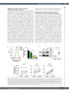
In mammals, mature red blood cells are devoid of mito- chondria. However, mitochondria can still be found at the reticulocyte stage in many mammalian species such as rabbits, dogs, rats, mice and humans.5,31-33 As expected, we detected a progressive decrease in mitochondrial biomass during erythroblast maturation, with a first major decrease in the transition between basophilic and poly- chromatic stages and a second between polychromatic and orthochromatic stages (Figure 3A). At day 10 of dif- ferentiation the mitochondrial marker TOM40 was detected at significantly higher levels in shVDAC1-trans- duced cells by western blot analysis (2.6±0.6-fold, n=5; Figure 3B). As VDAC1 downregulation alters the kinetics of differentiation, difference on protein expression at day 10 could results from a different proportion of erythrob- lastic stages. For this reason, mitochondrial biomass was assessed by flow cytometry in the different stages based on a-4integrin/Band3 profile as previously described. Mitochondrial biomass was significantly higher in shVDAC1-polychromatic and orthochromatic erythrob- lasts as compared to shSCR-transduced erythroblasts (Figure 3C). Importantly, these differences were associat- ed with late stages of differentiation as there was no change in mitochondrial biomass in earlier basophilic ery- throblasts. Although shSCR transduced cells progressive- ly eliminate mitochondria at each differentiation stage, our data point to a defect in mitochondrial clearance dur-
ing the transition between basophilic and polychromatic erythroblasts in conditions of VDAC1 downregulation.
VDAC1 regulates mitochondrial morphology and oxidative phosphorylation in terminal erythroblasts
In order to evaluate mitochondrial function in shVDAC1-transduced erythroblasts, we first evaluated the structural morphology of mitochondria by transmis- sion electron microscopy at day 10 of differentiation (Figures 4A; Online Supplementary Figure S3). In contrast with the normal ultrastructural morphology detected in shSCR-transduced cells, shVDAC1-transduced cells exhibited swollen mitochondrial cristae (Figure 4A and B) with a significantly increased percentage of rounded as compared to elongated mitochondria (P<0.0001, Figure 4A to C). These data are in agreement with a pub- lished role of VDAC1 OMM-IMM contacts sites in cristae structuration.23 Interestingly, we also observed a reduced number of ER-mitochondria contact sites in shVDAC1-transduced cells (Figure 4D). As these mito- chondrial associated membranes have been shown to recruit signaling molecules such as mTOR, GSK3 and hexokinase I, thereby increasing oxidative phosphoryla- tion,34 it was of interest to evaluate mitochondrial func- tion in erythroblasts with downregulated VDAC1. Notably, shVDAC1-transduced erythroblasts exhibited a significantly decreased oxygen consumption rate, evalu- ated as a function of mitochondrial biomass (Figure 4E). This measure, quantifying oxidative phosphorylation
AB
C
Figure 3. VDAC1 downregulation causes a retention of mitochondria in late stage erythroblasts. (A) Mitochondrial mass was measured by Mitofluor staining in ery- throblasts at different stages of terminal differentiation and representative histograms (left) and quantification of the mean fluorescence intensity (MFI) are shown (n=5) (right). (B) Levels of the mitochondrial protein TOM40 were quantified by immunoblot at day 10 of differentiation and representative blots (left) as well as nor- malization to b-actin (right) are presented. Levels in control cells was arbitrarily set at “1” (n=5). (C) Mitochondrial content of erythroblasts at day 11 of differentiation was evaluated by Mitofluor staining on basophilic (BasoE), polychromatic (PolyE) and orthochromatic (OrthoE) erythroblasts and MFI in progenitors transduced with scramble shRNA (shSCR) (black dots) and VDAC1 shRNA (shVDAC1) (red dots) transduced cells are presented (n=5). Gates for erythroblasts populations are based on a4-integrin/Band3 profiles as shown in Figure 1D. *P<0.05, **P<0.01. ProE: proerythroblast.
haematologica | 2022; 107(1)
171
