
Page 153 - 2022_01-Haematologica-web
P. 153
Oncogenic microRNA in T prolymphocytic leukemia
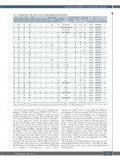
Table 1. Characteristics of the cohort of 31 T-cell prolymphocytic leukemia patients.
No Sex
1 M 2 M 3 M 4 F 5 F 7 M 8 M 9 F 10 M 11 M 12 M 13 F 14 M 15 M 16 M 17 F 18 M 19 F 20 M 21 M 22 F 23 F 24 M 25 M 26 F 28 M 29 M 44 M 46 F 48 F
49 M
Age WBC
Physical examination Survival Karyotype
FISH Clonality Immunophenotype 17p/ 11q / 14q/ TRB/TRG CD4/CD8 cyTCL1
L H
S Other Major abnormalities
65 740
58 670
41 79
63 393
50 14
74 42
60 69
77 11
789
79 55
49 34
67 115
62 245
67 59
62 643
77 72
61 61
67 493
69 28 n.a. n.a. n.a. n.a. n.a.
TP53 ATM TCL1
n.d. n.d. N n.d. n.d. n.d. n.d. n.d. n.d.
N N n.d. + ++ N N n.d. N NN
n.d. ++ N + n.d. N ++ + ++ N N+ N ++ + N+ + N n.d. N ++ N + n.d. N N n.d. N +N + ++ N N n.d. N + +
n.d. n.d. n.d. N + n.d. N++
n.d. n.d. n.d.
n.d. n.d. n.d.
n.d. n.d. n.d.
n.d. n.d. n.d.
clonal clonal clonal clonal clonal clonal clonal clonal clonal clonal clonal clonal clonal clonal clonal clonal clonal clonal clonal clonal clonal clonal clonal clonal clonal clonal clonal clonal clonal clonal clonal
CD4+CD8– + CD4+CD8– + CD4-CD8+ - CD4+CD8– + CD4+CD8+ + CD4+CD8– + CD4+CD8+ + CD4+CD8– - CD4-CD8- + CD4+CD8+ - CD4+CD8+ + CD4+CD8+ - CD4+CD8– - CD4+CD8– - CD4+CD8+ + CD4+CD8– + CD4+CD8– - CD4+CD8– + CD4+CD8– - CD4+CD8– + CD4+CD8+ - CD4+CD8– + CD4+CD8– - CD4+CD8+ + CD4–CD8+ - CD4–CD8+ + CD4+CD8+ + CD4+CD8– n.d. CD4+CD8– + CD4+CD8– + CD4+CD8– +
- -
- + + + + + - + + - - - - - - - + + + + +
n.d. n.d. n.d.
+ Sk 0.2 inv(14)(q32)
- 9 inv(14)(q32)/t(8;8) N + +
- 9 idic(8)(p11) - 0.5 inv(14)(q32) - 14 n.d.
- 28 n.d. -7n.d.
+ - + + + + + + + + - - - + + - + - - +
- 3.5 none -CNS7n.d. +-4n.d. +Sk87n.d. +-6n.d.
+- 5 none --26n.d. +Sk,L8n.d. +-10n.d. + Sk 47+ t(6;12)
+P16+
n.d. n.d. n.d. n.d. n.d. n.d.
69 24
72 231
89 550
67 25
77 243
48 31
44 27
52 50
- - -
+- +
- - - - - - + + - + + + + + + + - -
- n.a.
-8
-9
- 130+
- 4.5 inv(14)(q32)
- 7.7 -2+ - 3.5+
n.d. none n.d. inv(14)(q32) n.d.
81 120 n.a. n.a. n.a. n.a. dead
76 232 + - + - dead
77 293 n.a. n.a. n.a. n.a. n.a. inv(14)(q32)
75 48 - + - - dead n.d.
WBC:white blood cell count 109/L as determined at diagnosis;L:lymphadenopathy;H:hepatomegaly;S:splenomegaly;O:other; Sk:skin;L:lungs; P:pleural,A:ascites,CNS:cen- tral nervous system; TP53: TP53 abnormalities as detected by fluorescence in situ hybridization (FISH); ATM: ATM abnormalities as detected by FISH; TCL1: 14q32 translocations involving TCL1 as detected by FISH; cyTCL1: cytoplasmic TCL1 protein expression; n.a.: not available; n.d.: not determined; N: normal.
sorted CD4+ naïve (CD4N), CD4+ effector (CD4E), CD4+ memory (CD4M), CD8+ naïve (CD8N), CD8+ effector (CD8E) and CD8+ memory (CD8M) T-cell fractions from healthy individuals as reference. When using only T-PLL cases in a multidimensional APS plot analysis, the major- ity of T-PLL appeared to cluster according to similarities in their phenotype (Online Supplementary Figure S1B). However, two outliers were noted, T-PLL-10 and T-PLL- 24. Judging by the markers that were found to contribute to PC1 (mainly CD45RA) and PC2 (mainly CD45RA, CD45RO, CD27), the larger T-PLL cluster presumably would have a memory phenotype, while the outliers would resemble the naïve and/or effector T-cell subsets more (Online Supplementary Figure S1B). APS-based analy- sis of T-PLL cases that were added to fixed APS plots of normal T-cell subsets indeed confirmed that the majority of T-PLL cases did cluster close to or within the CD4+ and CD8+ memory T-cell subsets, whereas some T-PLL were more similar to non-memory subsets (Online
Supplementary Figure S1C). As clonal TRA and TRB gene rearrangements with stereotyped CDR3 have proven to be relevant for some T-cell leukemias, e.g., T-large granu- lar lymphocyte leukemia (T-LGL),25,26 we wondered whether this could be relevant for T-PLL as well. First, we asked whether V and J gene usage would define any sub- groups of the T-PLL samples. To this end, we performed Sanger sequencing of the clonal TRA and TRB gene rearrangements. In agreement with previously published data,27 sequence analysis of T-PLL samples revealed no clear skewing of V and J gene usage of TRA and TRB genes (Online Supplementary Figure S1D and E). In addi- tion, we did not find stereotyped CDR3 motifs in the pro- ductive TRA and TRB gene rearrangements in T-PLL either (data not shown).
In summary, most T-PLL displayed cell morphology and immunophenotypical characteristics reflecting normal memory T cells, with no clear evidence for antigen-driven leukemogenesis.
haematologica | 2022; 107(1)
145
