
Page 76 - 2021_12-Haematologica-web
P. 76
R. Hleihel et al.
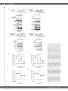
AB
CD
EF
Figure 4. Retinoic acid and chemother- apy cooperate to clear NPM-1c-express- ing cells in vivo. (A, B) Eight-week-old NSG mice were injected with 2x10⁶ OCI- AML3 cells intravenously. At day 21 after injection of the leukemic cells, retinoic acid (RA) was administered on a daily basis at the dose of 2.5 mg/g over a period of 1 week, followed by the administration of doxorubicin (2 mg/g) or cytarabine (AraC) (60 mg/g) twice a week over a period of 1 week. Mice were sacrificed, Bone marrow was harvested from femura and tibiae of xenografted mice and then stained with anti-human CD45 (hCD45) antibody. Western blot of NPM-1c, human P53, human P-P53 and PML in sorted hCD45-positive cells from BM harvested from untreated or treated NSG xenografted mice as indicated (2 mice per condition). (C, D) Eight-week- old NSG mice were intravenously inject- ed with 2x10⁶ OCI-AML3pml-/- cells. The same treatment regimen was followed as indicated above. Western blot of NPM-1c, human P53 and P-P53 in sort- ed hCD45-positive cells from bone mar- row harvested from untreated or treated
GH NSG xenografted mice as indicated. (E, F) Graphs showing the percentage of hCD45 cells in OCI-AML3 xenografted NSG mice treated as described above (7 mice in the untreated group and in the groups treated with doxorubicin alone or RA in combination with cytarabine [AraC], 9 mice in the group treated with RA alone or doxorubicin and RA, 8 mice in the group treated with cytarabine alone). (G, H) Graphs showing the per- centage of hCD45 cells in OCI-AML3pml-/- xenografted NSG animals treated as described above (3 mice in each condi-
tion).
3096
haematologica | 2021; 106(12)
