
Page 77 - 2021_12-Haematologica-web
P. 77
RA/arsenic activate a PML/P53 axis in NPM-1c AML
A
B
C
D
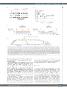
Figure 5. Retinoic acid and arsenic cooperate to clear NPM-1c-expressing cells. (A) Eight-week-old NSG mice were injected with 3x10⁶ OCI-AML3 or OCI-AML3pml-/- cells intravenously. From day 7 after injection of the leukemic cells, arsenic trioxide (ATO, 5 mg/g/day) and retinoic acid (RA, 2.5 mg/g) were administered intraperi- toneally every other day, over a period of 4 weeks. Western blot of human P53 and NPM-1c in sorted hCD45 positive bone marrow (BM) cells from NSG mice xenografted with OCI-AML3 or OCI-AML3pml-/- cells, after in vivo treatment with ATO alone, RA alone or the combination of RA and ATO. (B) Eight-week-old NSG mice were injected with 3x10⁶ OCI-AML3 or OCI-AML3pml-/- cells intravenously. From day 7 after injection of the leukemic cells, ATO and RA were administered every other day, over a period of 4 weeks intraperitoneally. At the end of treatment, bone marrow was harvested from femora and tibiae of xenografted mice and then stained with anti-hCD45 antibody. Graphs show the percentage of hCD45 cells in xenografted animals. (C) Treatment schedule in two patients with NPM-1c mutated acute myeloid leukemia (AML) treated with RA and ATO as indicated. Percentages of peripheral blood (PB) and bone marrow (BM) blasts are displayed. (D) Proposed model of the molecular mechanisms of the response of NPM-1c AML to RA. ITD: internal tandem duplication; NB: nuclear body.
The combination of retinoic acid and arsenic trioxide has clinical activity in mice and patients with acute myeloid leukemia
RA and ATO trigger NPM-1c degradation, P53 activation and apoptosis in NPM-1c-expressing AML cell lines.17,18 RA upregulates PML, while ATO targets PML to enforce nuclear body formation and also inhibits Pin1.6 Thus, in principle, ATO could cooperate with RA through PML nuclear body targeting.19 To explore any in vivo relevance of these findings, we treated xenografts from OCI-AML3 or OCI-AML3pml-/- with RA and ATO for 1 week. RA/ATO induced PML-dependent NPM-1c degradation and human P53 stabilization in vivo (Figure 5A) and decreased abun- dance of human cells in treated mice, again solely in cells bearing intact PML (Figure 5B).
The combination of RA/ATO is a very well-tolerated therapeutic association in APL.40 Two NPM-1c AML patients, unfit for conventional therapy, received this RA/ATO combination on an off-label compassionate basis. Blast clearance from peripheral blood and, to a lesser extent, from bone marrow was observed, although complete
remission was not achieved (Figure 5C). Longer follow-up after 2 months showed appearance of slowly growing AML cells. Thus, the RA/ATO combination may transiently tar- get AML cells in some NPM-1c-mutated patients.
Discussion
The basis for sensitivity of non-APL AML to RA was ini- tially believed to be RA-induced differentiation.11,41 Here we report that PML constitutes an unsuspected actor downstream of RA and is required for its synergistic activ- ity with other therapies in NPM-1c AML models. Previous ex vivo studies suggested that RA-driven NPM-1c degrada- tion could be the molecular basis of the therapeutic activ- ity of this drug through upregulation of ARF and resulting activation of P53. NPM-1c degradation should also correct multiple other phenotypes associated with NPM-1c, including sequestration of key regulators in the cytoplasm or transcriptional deregulation.4,42-45 Here, kinetic analysis of P53 activation upon RA treatment revealed that P53
haematologica | 2021; 106(12)
3097
