
Page 74 - 2021_12-Haematologica-web
P. 74
R. Hleihel et al.
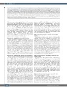
Figure 2. Retinoic acid targets PML/P53 through Pin1 inactivation. (A) Confocal microscopy of PML-nuclear bodies (NB) in primary blasts derived from one repre- sentative acute myeloid leukemia (AML) patient with wild-type (wt) NPM-1 and one representative AML patient with mutated NPM-1c, after ex-vivo treatment with retinoic acid (RA) or AF-17724 for 2 h as indicated. Histograms represente average number of PML NB per cell in two patients with NPM-1 wt and two patients with mutated NPM-1c. Statistical analysis was done using a Student t-test. (B) Western blot analysis of PML, P53 and total NPM-1 (NPM1-wt) in primary blasts derived from three patients with NPM-1c AML and three patients with NPM-1 wt AML, after ex-vivo treatment with 20 mM of AG17724 or 1 mM of RA for 2 h as indicated. (C) Western blot of PML and P53 in OCI-AML3 and OCI-AML2 cells following treatment with 20 mM of AG17724 for 2 h. Densitometry histograms represent an average of three independent experiments. (D) Colony-formation assays in methylcellulose of OCI-AML3, and OCI-AML3 Pin1-knock down (KD) cells, pre-treated with RA or AG17724 for 3 h (n=3). (E) Western blot analysis of PML, P53, P21, or Pin1 in OCI-AML3 and OCI-AML3 Pin1-KD after treatment with 1 mM of RA for 2 h. Densitometry histograms represent an average of three independent experiments. Densitometry was performed using ImageJ software. Statistical analysis was done using a Student t-test. (F) Western blot analysis of PML and P53 in OCI-AML3, OCI-AML3pml-/- and OCI-AML3P53-/- cells after treatment with 20 mM of AG17724 or 1 mM of RA for 2 h as indicated. Densitometry histograms represent an average of three independent experiments. Densitometry was performed using ImageJ software. Statistical analysis was done using a Student t-test, *P<0.05; **P<0.01; ***P<0.001.
When assessing the transcriptional effects of RA treatment in OCI-AML3 and control OCI-AML2 cells, a clear P53 sig- nature was noted in OCI-AML3 cells (Online Supplementary Figure S1C-E), in line with RA-induced P53 stabilization.17,18 To assess any role of P53 in cell death upon RA exposure, we also generated CRISPR P53 OCI-AML3 (OCI- AML3P53-/-) cell lines (Figure 1A, Online Supplementary Figure S1F). In this model, RA again failed to initiate cell death (Figure 1B, Online Supplementary Figure S1G), although it efficiently degraded NPM-1c (Figure 1A, Online Supplementary Figure S1F). Collectively, the RA-triggered, PML-facilitated NPM-1c degradation likely explains P53 activation and growth arrest of AML cells.
Retinoic acid targets P53 prior to NPM-1c loss
Further investigating the kinetics of response to RA, we unexpectedly obtained evidence of rapid P53 stabilization prior to any significant NPM-1c loss, but with a requirement for PML expression (Figure 1C). Thus, NPM-1c loss is not the sole contributor to P53 activation. Remarkably, similar data were obtained upon ex vivo treatment of primary blasts derived from NPM-1c AML patients, in whom RA-triggered NPM-1c loss was only obtained after 48 h, while P53 stabi- lization was generally observed as soon as 2 h (Figure 1D, E). Critically, such RA-triggered P53 activation was solely observed in samples from patinets with NPM-1c AML (Figure 1D, E). Thus, delayed NPM-1c degradation is most unlikely to explain early P53 activation upon RA treatment.
Retinoic acid stabilizes PML through Pin1 inactivation
RA also rapidly stabilized PML levels and induced PML nuclear body formation, solely in NPM-1c-positive patients' blasts or OCI-AML3 cells, with kinetics closely resembling those of P53 stabilization (Figure 2A, Online Supplementary Figure S2A-C). RA inconsistently enhanced PML gene expression, possibly through increased interferon produc- tion.35 Previous studies have shown that RA inhibits the Pin1 enzyme and the latter regulates PML stability.5,30,36 We thus compared the effects of RA and a Pin1 inhibitor (AG17724) on PML abundance, nuclear body formation and P53 activation. Strikingly, RA or AG17724 similarly sta- bilized PML or P53 levels and promoted nuclear body for- mation in NPM-1c AML patients’ blasts and OCI-AML3 cells (Figure 2A-C, Online Supplementary Figure S2A-C). In contrast, NPM-1-WT AML cells were unresponsive to RA and AG17724 (Figure 2A-C, Online Supplementary Figure S2A). Pin1 inhibition did not initiate NPM-1c degradation (Online Supplementary Figure S2D, E), implying that RA- induced PML stabilization is necessary, but not sufficient, to induce NPM-1c catabolism. Functionally, RA and AG17724 lead similarly to loss of clonogenic activity of OCI-AML3 cells in methyl-cellulose (Figure 2D). For a direct demonstra- tion of the involvement of Pin1 in the response to RA, we
generated an OCI-AML3 cell line with stable Pin1 down- regulation by shRNA. Downregulation of Pin1 did not affect the viability of NPM-1c AML cells (Online Supplementary Figure S2F). Remarkably, PML and P53 or P21 activation by RA was abrogated following downregulation of Pin1 (Figure 2E), and RA-induced cell death was lost (Online Supplementary Figure 2F). Loss of clonogenic activity by RA or Pin1 inhibition was also abrogated (Figure 2D). Thus, RA-induced activation of P53 and resulting growth arrest are initiated by Pin1 inhibition.
PML is the primary target of retinoic acid and Pin1 inhibitors
Pin1 directly controls both the stability of PML and P53 signaling.37 PML and P53 are highly cross-regulated: PML controls P53 activation, but P53 transcriptionally induces PML expression.38,39 To decipher the respective roles of PML and P53 in response to Pin1, we compared the responses to RA and AG17724 in OCI-AML3 and its pml-/- and P53-/- derivatives. Both drugs upregulated PML levels in P53-/- cells, and no (or minimal) induction of P53 was observed in OCI- AML3pml-/- cells (Figure 2F). These results establish that PML is the primary target of Pin1 inhibition in NPM-1c-express- ing cells. These results support a model wherein RA inacti- vates Pin1, to stabilize PML, induce nuclear body forma- tion, activate P53 and suppress growth. This does not exclude the possibility that P53 then constitutes a feed-for- ward amplification loop on PML expression.
NPM-1c-expressing cells exhibit high basal level and activity of Pin1
The implication of Pin1 inhibition in RA-mediated effects in NPM-1c-expressing cells prompted us to investigate Pin1 levels and activity. Strikingly, high Pin1 protein levels and activity were observed in NPM-1c-expressing cell lines and primary blasts (Figure 3A-C, Online Supplementary Figure S3A). Treatment with RA significantly reduced Pin1 activity (Figure 3C) without affecting Pin1 protein level (Online Supplementary Figure S3B). In line with this increased Pin1 protein level and activity in NPM-1c-expressing cells, both RA and AG17724 synergized with arsenic trioxide (ATO), and standard chemotherapy drugs used in AML, doxoru- bicin or cytarabine, to induce death of OCI-AML3 cells but not of OCI-AML2 cells (Figure 3D).
Retinoic acid and chemotherapy cooperate to clear NPM-1c-expressing cells in vivo
The clinical benefit of co-administration of RA with chemotherapy seems restricted to AML bearing an NPM-1c mutation.16 We thus examined the possibility of in vivo cooperation between RA and standard-of-care AML thera- py, anthracyclins and cytarabine, and examined any PML dependency. We used xenografts from OCI-AML3 or OCI-
3094
haematologica | 2021; 106(12)
