
Page 72 - 2021_12-Haematologica-web
P. 72
R. Hleihel et al.
AB
C
D
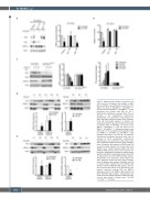
E Densitometry histograms represent data from an average of three independent experiments. Densitometry was performed using ImageJ soft- ware. Statistical analysis was done using a Student t-test. (D) Western blot analysis of NPM-1c and P53 in primary blasts derived from five patients with NPM-1c acute myeloid leukemia (AML) and three NPM-1wt AML patients, after ex-vivo treatment with RA for 2 h. Densitometry histograms represent aver- age P53 and NPM-1c expression levels in five NPM- 1c AML patients and three NPM-1wt AML patients. Densitometry was performed using ImageJ soft- ware. Statistical analysis was done using a Student t-test. (E) Western blot analysis of NPM-1c and P53 in primary blasts derived from five NPM-1c AML patients and three NPM-1wt AML patients, after ex- vivo treatment with RA for 48 h. Densitometry his- tograms represent average P53 and NPM-1c expression levels in five NPM-1c AML patients and three NPM-1wt AML patients. Densitometry was performed using ImageJ software. Statistical analy- sis was done using a Student t-test, *P<0.05;
Figure 1. PML-dependent NPM-1c degradation and P53 activation. (A) Western blot analysis of PML, P53 and NPM-1c was performed on extracts of OCI- AML3, one clone of OCI-AML3pml-/- (OCI-AML3pml-/-#1) and one clone of OCI-AML3P53-/- (OCI-AML3P53-/-#1) after treatment with retinoic acid (RA) for 48 h. Densitometry histograms represent data from an average of five independent experiments. Densitometry was performed using ImageJ soft- ware. Statistical analysis was done using a Student t-test. (B) Cell growth (percent of control) was assessed using the trypan blue exclusion dye assay, in triplicate wells in OCI-AML3, one clone of OCI- AML3pml-/- (OCI-AML3pml-/-#1) and one clone of OCI- AML3P53-/- (OCI-AML3P53-/-#1) following treatment with RA for 48 h (n=3). (C) Western blot analysis of P53 and NPM-1c in OCI-AML3 and OCI-AML3pml-/- after treatment with RA for 2, 12 and 24 h as indicated.
**P<0.01; ***P<0.001.
3092
haematologica | 2021; 106(12)
