
Page 30 - 2021_12-Haematologica-web
P. 30
Ž. Antić et al.
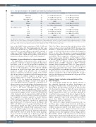
Table 1. Cox regression analysis in the combined representative ALL9 and ALL10 cohort (n=376).
IKZF1
MRD
Age at diagnosis, years
Gender
Status
High-clonal3 Subclonal Other high-clonal4 Low Medium High
0-4
5-9
10-14
15-18
Male
Female
Number of patients
18
22
13
111
227
22
173
133
47
23
215
161
Univariate Cox regression1,2
P<0.01; HR=7.22 [3.27-15.95]
P=0.39; HR=1.69 [0.51-5.57]
P<0.01; HR=19.92 [9.76-40.66]
1 (Ref)
P=0.1; HR=2 [0.88-4.61]
P<0.01; HR=10.85 [4.16-28.28]
1 (Ref)
P=0.34; HR =1.34 [0.73-2.46]
P=0.83; HR=0.9 [0.34-2.38]
P=0.3; HR=1.76 [0.6-5.12]
1 (Ref)
P=0.06; HR=0.56 [0.31-1.03]
Multivariate Cox regression1,2
P<0.01; HR=3.6 [1.38-9.55] P= 0.34; HR=1.8 [0.53-6.24] P<0.01; HR=13 [5.68-29.79] 1 (Ref)
P=0.28; HR=1.6 [0.67-3.78] P<0.01; HR=5 [1.64-15.23] 1 (Ref)
P=0.6; HR=1.2 [0.64-2.25] P=0.6; HR=0.8 [0.27-2.12] P=0.3; HR=0.34 [0.04-2.66] 1 (Ref)
P=0.08; HR=0.53 [0.26-1.1]
1The hazard ratio is given with a 95% confidence interval.Multivariate Cox regression analysis included gender,age at diagnosis and minimal residual disease status as covariates; 2Analysis was done on the combined cohort stratified on treatment protocol. Representative ALL9 and ALL10 cohorts are outlined in Online Supplementary Table S1. 3Clonality status based on detection at initial diagnosis; high-clonal: allele frequency ≥25%, subclonal: allele frequency <25%. 4Detectable using a multiplex ligation-dependent probe ampli- fication assay. A full overview of all comparisons is given in Online Supplementary Table S11. MRD: minimal residual disease.
that of the NRAS hotspot mutations G13D, G12D and KRAS A146V (Figure 3E). This finding indicates that some RAS hotspot mutations (e.g., KRAS G12D, G13D, A146T) may result in a stronger expansion potential compared to others (e.g., KRAS A146V, NRAS G12D, G13D), and fur- ther illustrates the complex heterogeneity of RAS hotspot mutations in their potential to drive clonal expansion.
Relevance of gene alterations to relapse development
The high number of alterations in these relapse-associ- ated genes at the time of diagnosis triggers the hypothesis that these could be used as prognostic biomarkers for relapse development, even when present at subclonal lev- els. To test this hypothesis, we first explored whether alterations in each of the eight genes were enriched in diagnostic samples from patients who subsequently relapsed compared to diagnostic samples from patients who did not relapse. In general, subclonal alterations were very common at primary diagnosis in patients who relapsed (60/82; 73%) as well as in patients who did not (165/203; 81%). For high-clonal alterations, we only observed a higher percentage of relapse development in cases with IKZF1 deletions compared to wild-type cases, whereas an association with relapse development was not observed for diagnostic samples with subclonal alterations in any of the genes, including IKZF1 (Figure 4). Furthermore, patients with high-clonal IKZF1 4-7 dele- tions were more often classified as having high minimal residual disease (MRD; >5×10⁻⁴ at day 79 or 84 after start of the treatment) in both representative ALL9 and ALL10 cohorts (Fisher exact test, P<0.01 and P<0.05, respective- ly), compared to patients without an IKZF1 deletion (Online Supplementary Table S10). The CIR at 5 years was 41.7% (SE 0.04%) and 42.9% (SE 0.03%) in patients with high-clonal IKZF1 4-7 deletions treated according to the ALL9 and ALL10 protocols, respectively (Figure 5). The cause-specific hazard ratio (HRCS) in the two representa- tive cohorts (n=376), estimated with a univariate Cox pro- portional hazards regression model, revealed an associa- tion of high-clonal IKZF1 exon 4-7 deletions with relapse (HR=7.22; 95% CI: 3.27-15.95; P<0.01). In the multivari- ate Cox model, in which age at diagnosis, gender and MRD status were included, the adjusted HRCS was 3.6 (95% CI: 1.38-9.55; P<0.01) (Table 1, Online Supplementary
Table S11). These data are in line with those from earlier studies on these cohorts in which all IKZF1 deletions were included.12,24 However, when we assessed the clinical rel- evance of subclonal alterations for relapse development in IKZF1, or any of the other genes, Cox regression analysis revealed no significant associations in the combined ALL9 and ALL10 cohorts compared to wild-type cases (Table 1, Online Supplementary Table S11), and the CIR was similar in the two groups (Figure 5). Furthermore, patients with subclonal IKZF1 4-7 deletions did not have significantly different levels of MRD compared to IKZF1 wild-type patients (Online Supplementary Table S10). Since previous studies have shown a lack of association of IKZF1 deletion with relapse in patients who carry a deletion in ERG,34,35 we used MLPA to test whether there was an enrichment of ERG deletions in cases with subclonal IKZF1 exon 4-7 deletions compared to those with clonal IKZF1 exon 4-7, but these deletions were infrequent in both groups (Online Supplementary Table S12).
Tracing of major and minor clone mutations at the time of relapse
To obtain further insight into the clinical relevance of the identified alterations in relapse development, we investigated whether these were preserved in the cases that relapsed. For this analysis, we used all 146 cases that later developed a relapse, of which 82 carried alterations in a major or minor clone in one or more of the genes (Online Supplementary Tables S13 and S14). Overall, we found that for most genes at the time of diagnosis the fre- quency of subclonal alterations was similar or slightly higher compared to that of the alterations detected in a major clone (Online Supplementary Figure S3A, Online Supplementary Tables S13 and S14).
We collected 73 relapse samples from patients who car- ried these major or minor clone alterations at the time of diagnosis (89%), which enabled us to trace 171 of the 185 sequence mutations, and 25 of the IKZF1 exon 4-7 dele- tions. We did not assess whether mutations detected at diagnosis were still preserved in minor clones at relapse, since these clones were unlikely to be true relapse drivers. Overall, 56% (22/39) of the tested major clone mutations were found to be preserved in the major clone at relapse, whereas the value for the subclonal mutations was 7%
3050
haematologica | 2021; 106(12)
