
Page 122 - 2021_12-Haematologica-web
P. 122
S. Bohler et al.
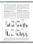
there were no differences between the viruses used (Figure 4A). As known from this model system, most cells differ- entiated into CD19+ B cells while fewer CD33+ myeloid cells and almost no CD3+ T cells arose (Figure 4B and data not shown). Cells expressing the control shRNA (Luci) engrafted and contributed to all lineages (Figure 4C, D). In contrast, cells expressing shRNA specific for MCL-1 showed only very poor engraftment (Figure 4C). In line with the important role of MCL-1 for survival of immature progenitors with multipotent potential, all cell types found in the xenografted mice were equally affected (Figure 4D).
MCL-1 inhibition limits the survival of immature but not mature hematopoietic cells
To determine the effects of MCL-1 inhibition on more mature types of hematologic cells, we used the specific MCL-1 inhibitor S63845. First, we treated freshly isolated immature CD34+ and mature CD34- cells with increasing doses of the inhibitor. While CD34+ cells were moderately sensitive when compared to cancer cell lines (e.g., IC50 in most multiple myeloma cell lines <0.1 mM),28 no apoptosis was induced after 24 h and 48 h in mature CD34- blood cells even when very high doses of inhibitor were used (Figure 5A, B).
In a second approach, we let untreated CD34+ cells dif- ferentiate for 11 days in MethoCult medium. Cells were
AB
then isolated and put into stem cell medium containing 10% ES-FBS and cytokines (SCF, TPO, FLT3L, IL-3). Different concentrations of the MCL-1 inhibitor were added, and cell numbers were determined after 24 h. Again, differentiated CD34- cells were much less sensitive than immature CD34+ cells (Figure 5C, D). As a conse- quence, only a mild and non-significant reduction in cell numbers was noted (Figure 5C) and both myeloid and ery- throid cells were depleted only to a minor and non-signifi- cant degree (Figure 5D).
Stem and progenitor cells from neonates and adults are equally sensitive to MCL-1 or BCL-XL inhibition
Our experiments indicate a strong dependence of human HSPC on MCL-1 expression, which is not unexpected con- sidering the high relevance of MCL-1 for survival of murine HSPC.25,26 However, other authors described an overall good tolerability of human HSPC to MCL-1 inhibitors.33-35 One reason for this discrepancy could be that we used CD34+ cells derived from cord blood while CD34+ cells derived from the bone marrow of aged persons were used in other studies.33 We therefore compared these two cell types with regards to protein levels of MCL-1 and other anti-apoptotic BCL-2 family members. Bone marrow CD34+ cells were obtained from patients with orthopedic problems (age range: 44 to 90 years). While MCL-1 and
CD
Figure 4. Human CD34+ cells lacking MCL-1 show poor engraftment in xenografted mice. (A-D) Lentivirally transduced or untransduced human hematopoietic stem and progenitor cells (HSPC) were transplanted intrahepatically into newborn Rag2−/−gc−/− mice after sub-lethal irradiation. Mice were sacrificed 8 weeks after trans- plantation and bone marrow (BM) and spleen populations were analyzed. By using antibodies specific for human or murine CD45, percentage human engraftment was determined (A). The various human hematopoietic populations were determined within the huCD45+ cells using flow cytometry (B). GFP expression was deter- mined in huCD45+ cells (C) and in each of the subpopulations (D). Bars represent mean ± standard error of mean, n =9-13 from eight independent experiments. Mann–Whitney test was performed; *P<0.05, **P<0.01.
3142
haematologica | 2021; 106(12)
