
Page 120 - 2021_12-Haematologica-web
P. 120
S. Bohler et al.
either MCL-1 (shown here) or BCL-XL (documented ear- lier)31 are further reduced by RNA interference.
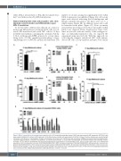
Human hematopoietic stem and progenitor cells show impaired colony formation and differentiation upon MCL-1 knockdown
To test the effect of MCL-1 knockdown on colony for- mation and differentiation of human CD34+ cells, we cul- tured 1,000 transduced and sorted GFP+ cells for 11 days in MethoCult medium containing the cytokines SCF, IL- 3, IL-6, erythropoietin (EPO), granulocyte colony-stimu- lating factor (G-CSF), granulocyte-macrophage colony- stimulating factor (GM-CSF), insulin and transferrin. The
number of colonies arising was significantly lower when MCL-1 expression was inhibited (Figure 2A). All colony types were affected, indicating that all multipotent and lineage-committed progenitor cell types were lost to a similar degree (Figure 2B). In addition, fewer cells could be harvested from plates (Figure 2C). Flow cytometry revealed that all cell types were reduced in number when MCL-1 was depleted (Online Supplementary Figure S3A). Since we noticed a relevant toxicity of the sorting proce- dure on lentivirally-transduced cells, we repeated the experiment using unsorted cells. With this approach, we could directly compare transduced GFP+ with untrans- duced GFP- cells. Colony numbers and types were similar
ABC
DEF
G
Figure 2. MCL-1 is essential for all hematopoietic progenitor cells. (A-C) Lentivirally transduced human CD34+ cells were sorted for GFP expression. GFP+CD34+ cells were seeded in MethoCult medium (1,000 cells each). (D-G) Alternatively, unsorted cells were plated. (A, D) After 11 days of culture, colonies were counted by light microscopy. (B, E) Based on morphological findings, the following colony types were identified by light microscopy: GEMM: granulocytic-erythroid-megakaryocytic- monocytic, GM: granulocytic–monocytic, G: granulocytic, E: erythroid, M: monocytic. (C, F) Cells were dissolved from the semisolid medium and counted by a hemo- cytometer. (G) The different hematopoietic cell types were determined by flow cytometry. The percentages of GFP+ cells are shown within each of the following cell populations: HSC; hematopoietic stem cells (CD34+CD38-CD45RA-CD90+), MPP: multipotent progenitors (CD34+38–CD45RA-CD90-), GM: granulocytic–monocytic pro- genitors (CD34+CD33+CD115+), CFU-G: colony forming unit-granulocytes (CD34-CD33+CD15+CD115-), M: monocytes (CD34-CD33+CD14+CD115-), immE: immature ery- throcytes (CD71hiCD235a-), matE: mature erythrocytes (CD71+CD235a+). Bars represent mean ± standard error of mean, n=6 (A-C) from six independent experiments and n=4 (D-G) from four independent experiments. Mann-Whitney test: *P<0.05, **P<0.01.
3140
haematologica | 2021; 106(12)
