
Page 121 - 2021_12-Haematologica-web
P. 121
MCL-1 in human hematopoiesis
in all groups (Figure 2E, F) and cell numbers were not con- sistently reduced when MCL-1 was downregulated (Figure 2G). However, while transduction rates were comparable (Online Supplementary Figure S3B), MCL-1- depleted GFP+ cells were selectively lost during the 11 days of MethoCult culture, indicating their selective dis- advantage (Figure 2H, Online Supplementary Figure S3B). Loss of MCL-1 affected all analyzed immature and mature cell types in a similar manner, confirming that all progenitor cell types were dependent on MCL-1 expres- sion (Figure 2H, Online Supplementary Figure S3C). To ana- lyze whether MCL-1-depleted cells were lost immediate- ly or progressively over time, we cultured them for only 5 days in MethoCult medium. At this early time point, only mild loss of GFP+ cells was observed, independently of the lentivirus used (Figure 3A, left). This indicates that progenitor cells became more susceptible to MCL-1 inhi- bition once they progressed in their differentiation process. Accordingly, immature CD34+ cells and specifi- cally hematopoietic stem cells and multipotent progeni- tors were enriched in the first days of culture (Figure 3B, Online Supplementary Figure S3D, E). To inhibit differenti- ation but foster proliferation, CD34+ cells were cultured in the presence of the cytokines SCF (200 ng/mL), FLT3L (200 ng/mL), TPO (100 ng/mL) and IL-3 (20 ng/mL). CD34 and GFP expression was measured after 5 and 11 days. Under this condition, the percentage of CD34+ cells remained high (Figure 3B, right) and GFP+ cells were not depleted in a relevant manner (Figure 3A, right). To exclude that expression of shRNA and, consequently, MCL-1 knockdown was different in the two culture con- ditions, we measured MCL-1 mRNA after 5 days of cul- ture. While we observed stable MCL-1 knockdown by shRNA #3, the shRNA #4 showed less consistent results
with re-expression of MCL-1 mRNA (Online Supplementary Figure S3F). However, no difference in knockdown efficiency was observed between the two culture conditions indicating that the dependence of HSPC on MCL-1 did indeed change during proliferation and differentiation, respectively.
To understand why MCL-1 dependence was so different under the two culture conditions, we determined the com- position of all BCL-2 proteins by RT-MLPA. As controls, we used freshly isolated CD34+ cells. Interestingly, both MCL-1 and BCL-XL were expressed at higher levels in cells stimulated to differentiate for 4 days (Figure 3C). Among the pro-apoptotic BCL-2 proteins, PUMA was highly upregulated under both culture conditions (i.e., differentia- tion and proliferation conditions) while BIM, BID and BAK1 were selectively upregulated under differentiation conditions (Figure 3D, E). Thus, it is possible that differen- tiation is associated with stronger pro-apoptotic signals that need to be counteracted by higher MCL-1 and BCL- XL levels. Based on its binding affinities, it is conceivable that MCL-1 expression is required to counteract BIM- mediated activation of BAK1.
MCL-1 inhibition severely restricts hematopoietic stem andprogenitorcellengraftmentinxenograftedmice
In order to determine the effects of MCL-1 inhibition on the engraftment potential of human CD34+ HSPC, untrans- duced and transduced cells were intrahepatically trans- planted into sublethally irradiated Rag2-/-gc-/- mice. To reduce cell stress prior to transplantation, we waived the sorting procedure and transplanted GFP+ cells together with GFP- cells. After 8 weeks, xenografted mice were sac- rificed and human engraftment was analyzed. Lentivirally transduced cells had a reduced potential to engraft but
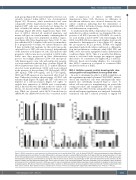
AB
CDE
Figure 3. MCL-1 expression is more important for differentiating than for proliferating CD34+ cells. (A) Sorted CD34+GFP+ cells were subjected to either differentiat- ing or proliferating culture conditions. To induce differentiation, cells were cultured in semisolid MethoCult plates for 5 days (3,000 cells seeded per plate) or 11 days (1,000 cells seeded per plate). To foster proliferation, cells were cultured in stem cell medium containing 10% serum and stem cell factor, FLT3L (200 ng/mL each), thrombopoietin (100 ng/mL) and interleukin-3 (20 ng/mL). After 5 and 10 days of culture, the percentages of GFP+ cells were determined by flow cytometry. (B) The fraction of CD34+ cells was determined within GFP+ cells at each time point. (C-E) Untransduced CD34+ cells were subjected to the two different culture conditions and harvested at the indicated time points. mRNA was used for reverse transcriptase-multiplex ligation dependent probe amplification designed to determine levels of apoptosis genes. Results are shown for anti-apoptotic BCL-2 proteins (C), BH3-only proteins (D) and pro-apoptotic effector proteins (E). Freshly isolated CD34+ cells were used as controls. Bars represent mean ± standard error of mean, n=4 from four independent experiments. Mann-Whitney test: *P<0.05, **P<0.01.
haematologica | 2021; 106(12)
3141
