
Page 201 - 2021_10-Haematologica-web
P. 201
PIMT and RBC metabolism
F
H
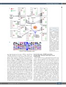
ing 6-phosphogluconolactonase (6PGL), transaldolase (TALDO), glutathione peroxidase 1 (GPX1) (Figure 1C). Conversely, the levels of other enzymes involved in alter- native redox regulation pathways were increased in PCMT1+/- RBC, including peroxiredoxin 2 (PRDX2), thiore- doxin reductase 2 (TRXR2), hydroxyacyl glutathione hydrolase (GLO2), glucose 6-phosphate isomerase (G6PI), lactate dehydrogenase B (LDHB) (Figure 1C). Accordingly, we focused on these pathways to provide a more detailed overview of the metabolic changes observed in RBC from these mice (Figure 1F). Specifically, we noted an accumula- tion of methionine (the main methyl group donor precur- sor - Figure 1F), consistent with a decreased PIMT levels and activity. Likewise, differential D-methylation patterns were observed between WT and PCMT1+/- RBC, especially in regions in which aspartyl groups are flanked by other negatively charged residues (D, E, S) in proximity of deamidation susceptible asparaginyl groups (N – represen- tative methylation of deamidated N is shown for the most abundant protein, hemoglobin - Figure 1G, the full list is stated in the Online Supplementary Table S1). Notably, sig- nificantly higher levels of band 3 methylation of residues D329 and 368 was observed in WT mice compared to het- erozygous mice (Online Supplementary Table S1).
Figure 1. Metabolomics and proteomics of heterozygous PCMT1+/- mouse red blood cells. (A) Metabolomics and proteomics of heterozygous PCMT1+/- (heterozygous
G PCMT1) mouse red blood cells (RBC); (B) and (C) top 50 metabolites and proteins by t- test between wild-type (WT) and heterozygous PCMT1 mice, respectively; (D to F) Partial Least Square- Discriminant Analysis, Variable Importance in Projection and methionine metabolism in RBC from WT and heterozygous PCMT1 mice; (G) quantitative analysis of methylated deamidated asparagine residues in hemo- globin – as representative for the RBC proteome; (H) pre- ferred protein methylation motifs as identified in the RBC proteome of WT vs. heterozy-
gous PCMT1 mice.
Lack of both copies of PCMT1 exacerbates
the phenotypes observed in heterozygous mouse red blood cells
PCMT1 KO mice die at approximately 6-8 weeks from seizures due to accumulated protein damage in brain tissue.15,26 PCMT1 KO mice were obtained by crossing of PCMT1 heterozygous mice and were characterized at 5 weeks of age (prior to the onset of seizures) compared to age-matched WT mice; to ensure that the same background genetics were present in each group, PCMT1 KO, WT and heterozygous mice were all obtained from the same colony of interbreeding heterozygous mice (Figure 2A). Unsupervised principal component analysis (PCA) revealed a progressive alteration of the metabolic phenotype of RBC from WT to heterozygous and KO mice (Figure 2B), with the main metabolic discriminants across groups being pen- tose phosphate pathway (PPP) metabolites (6-phosphoglu- conate, sedoheptulose phosphate, ribose phosphate), polyunsaturated fatty acids (eicosa- and docosahexaenoic acid), oxylipins (15-HETE), tryptophan and tyrosine metabolites (including several indoles, picolinic acid, dopamine – Figure 2C). Changes in the metabolome and proteome (Figure 2D to E) extended the observations from the heterozygous mouse RBC, showing an apparent gene
haematologica | 2021; 106(10)
2729
