
Page 43 - 2021_09-Haematologica-web
P. 43
Monitoring of clonal evolution of AML
AB
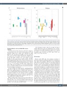
Figure 2. Variation of the allelic frequency of the variants. Box plots representing the increase or decrease of allelic frequencies detected in genes where variants have been detected at refractoriness. (A) and at relapse (B). The genes are grouped according to metabolic pathways, with different colors representing: transcrip- tional regulator genes (ASXL1, EZH2 and PHF6) in green, CALR in black, epigenetic regulator genes (DNMT3A, TET2, IDH1, IDH2, KDM6A and KMT2A) in yellow, splicing genes (SF1, SF3A1, SF3B1, SRSF2, U2AF1, ZRSR2 and PRPF40B) in brown, cytokine signaling and JAK/STAT pathway genes (EPOR, FLT3, JAK2, KIT, SH2B3, MPL and CBL) in blue, GTPase activity genes (HRAS, KRAS and NRAS) in pink, transcription factors genes (ETV6 and RUNX1) in grey and tumor suppressor genes (VHL, TP53 and PTEN) are represented in red.
Clonal evolution is not associated with a worse outcome
Conventional molecular alterations detected at diagnosis in ten patients were lost both at refractoriness (n=3) and leukemia relapse (n=7). AMA were identified in eight patients, who all achieved CR (eight of 17 cases with any CR vs. zero of six cases with no CR; P=0.037). Median over- all survival was 77.4 (range, 21.5–133.3) months for the group with AMA features versus 11.8 (range, 1.2–22.4) months for the group without AMA features (P=0.083, Online Supplementary Figure 3SA). Clonal evolution detected with AMA identified patients with a trend for a better prog- nosis for disease-free survival (median disease-free survival was 22.1 vs. 10.8 months; P=0.065, Online Supplementary Figure 3SB); however, neither loss of molecular abnormali- ties nor combined additional and lost molecular abnormali- ties had an impact on prognosis.
Accrual of AMA was mainly related to signaling pathway genes (five of eight cases cases with gain of mutations in the genes of the signaling pathway compared with only three of 15 cases without gain of mutations; P=0.042). Loss of AMA in relapsed samples was also mainly found for signaling pathway genes (seven of ten cases with loss of AMAs pres- ent in signaling pathways genes compared with one of 13 cases without loss of AMA; P=0.002).
Other variables with a trend for an association with cases who achieved CR were normal karyotype (six of 17 cases with any CR vs. zero of six cases with no CR; P=0.091) and cytogenetic risk (only six of ten high-risk, but 11 of 13 inter- mediate- or low-risk achieved CR; P=0.183).
New therapeutic targets, either by an approved drug or within clinical trials, were not identified in cases of refrac- toriness; however, eight potential new targets were found in five relapsed cases (zer of ten refractoriness cases vs. five of 13 relapse cases, P=0.027): one IDH2, three SF3B1, two KRAS, one KIT and one JAK2.
Discussion
Patients with AML who show primary resistance to induction treatment or leukemic relapse have a dismal prog- nosis. Our study identifies differences in the mutational landscape between primary refractory and relapsed AML. In this line, we report the usefulness of monitoring different leukemic clones, and particularly detecting the appearance of new clones, using an NGS-targeted panel in post-treat- ment AML, allowing us to: i) stratify patients into prognostic risk groups; ii) select MRD marker/s to monitor response to treatment; and ii) define targeted post-remission strategies including the selection of new drugs for leukemia relapse.
The genetic follow-up of leukemic clones was performed using NGS technology with a high coverage of the variants (>1,000×), allowing the detection of sub-clones with a high sensitivity (<3%) in a cohort of 23 patients with AML, and with almost 100 samples evaluated. The NGS technology also allowed us to estimate the mutational load based on the VAF level, and to, therefore, infer the clonal architecture of the tumor and the model of clonal evolution.6 We confirmed the high clonal heterogeneity associated with this disease and the mutational profile associated with treatment refrac-
haematologica | 2021; 106(9)
2331
