
Page 42 - 2021_09-Haematologica-web
P. 42
E. Onecha et al.
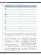
Table 2. Variants detected in refractoriness, complete remission and relapse versus diagnosis.
Refractoriness Relapse Complete Remission
Dx + Dx + Dx - Dx + Dx + Dx - Dx + Dx + Dx - Rf+ Rf- Rf+ R+ R- R+ CR+ CR- CR+
ASXL1 1 3 3
CALR 1
CBL 3 11 2
DNMT3A 61 64
EPOR 32 ETV6111 3 EZH2122 6 FLT3-SNV1 41 6
IDH1 2
IDH2 4 125
JAK2 121
KMD6A 1
KIT3 1
KMT2A 41111
KRAS 2
MPL 2 3
NRAS 3
PHF6 3
PRPF40B
PTEN
RUNX1 2
SF3A1
SF3B1
SRSF2
TET2 2
TP53 3112 1
U2AF1 2 1 3
VHL 1215
ZRSR2 1
N 21 10 5 42 10 18 28 53 5
The table lists the number of samples with allelic variants detected in the different genes included in the study.The table specifies the samples studied in primary refractoriness, relapse and complete remission.The last row indicates the number of samples that are present at diagnosis (Dx +),at refractoriness (Rf +),at relapse (R +) or at complete remis- sion (CR +).Variants that are present in the diagnosis (Dx +) but absent in refractoriness (Rf -) or relapse (R -) or in complete remission (CR-).Thus,there are variants absent in the diagnosis (Dx -) but are present in refractoriness (Rf +) or relapse (R +) or complete remission (CR +).
13 36 1 2 1
1
2 5
1 343
3
1
21 44
3
(n=4/8), EZH2 (n=6/6), RUNX1 (n=5/5), VHL (n=5/5), FLT3 (n=6/6), SF3B1 (n=4/6), ETV6 (n=3/3), U2AF1 (n=3/3), PHF6 (n=2/2), y KMT2A (n=1/2). Also, five variants arose de novo in DNMT3A (n=2) and MPL (n=3). These results provide potential markers that could be used to detect minimal residual disease (MRD) in our series, including EZH2, RUNX1, VHL, FLT3, ETV6, U2AF1, PHF6 and SF3B1, as these variants disappear in CR.
Branching clonal evolution is predominant in acute myeloid leukemia
Analysis of the molecular dynamics of the clones accord- ing to the VAF identified three patients who showed a change in the predominant clone from diagnosis to primary refractoriness: clones characterized by mutations in VHL (patient 1), ETV6 (patient 2) and TP53 (patient 6) became the predominant clones at refractoriness (Figure 1A). Likewise, four relapsed patients showed changes in the predominant clone from diagnosis, characterized by mutations in EPOR
(patient 9), TP53 (patient 11), VHL (patient 16) and PHF6 (patient 22) (Figure 1A to C). In addition, clonal evolution was observed in 12 patients. A linear clonal evolution model was identified in four patients in primary refractoriness and two in relapse. By contrast, a branching clonal evolution model was identified in two patients in primary refractori- ness and in six patients in relapse.
Subclonal mutations (VAF <10%) were detected at diag- nosis in signaling pathway genes (JAK2, FLT3, NRAS) and in splicing genes (U2AF1, SF3B1). Of these, JAK2, NRAS and U2AF1 variants disappeared in the treatment failure sam- ples, whereas FLT3 and SF3B1 variants persisted. Also, we detected variants in TP53 and PHF6 (tumor suppressor genes), but only the PHF6 variant became the predominant clone in a relapsed sample (VAF=83%). Two TP53-subclonal mutations were detected at diagnosis in the same patient, but only one of them was maintained at a similar frequency in the refractory sample.
2330
haematologica | 2021; 106(9)
