
Page 77 - 2021_06-Haematologica-web
P. 77
Hyaluronan/CD44-mediated VLA-4 activation in AML
(clone HP2/1) and intravenously injected into NSG mice. Mice were sacrificed after 3 h (short-term homing, allowing leukemia cell entry into organs but no proliferation), and transplanted cells were flow cytometrically identified in the spleen and BM by human-specific antibodies (Figure 1B). Cells that had been treated with the blocking αCD44 Fab fragment had a lower capacity to home to BM within 3 h compared to untreated cells. The homing of primary human AML cells to the spleen was strongly diminished upon CD44 blockade. In contrast, αCD49d antibody treat- ment only slightly reduced BM homing of AML blasts and had no effect on their spleen homing (Figure 1Ci). In four of the five homing experiments we combined αCD44/αCD49d treatment, but additional CD49d block- ade did not further increase the inhibitory effect above the level achieved by treatment with αCD44 alone (Online Supplementary Figure S1B). Comparable effects were observed when using OCI-AML3 cells (Figure 1Cii). The significantly reduced recovery of αCD44-treated cells was not due to toxicity of the antibody, as in vitro treatment for 3 h had no effect on cell viability (Online Supplementary Figure S2A). Neither the functional inhibition of CD44 nor the inhibition of CD49d affected the general CD44 and CD49d expression of the cells (Online Supplementary Figure S2B). Anti-CD44 antibody treatment did not affect CD44/E-selectin-mediated cell arrest (Online Supplementary Figure S2C). We genetically confirmed the contribution of CD44 to homing by CD44 knockdown in OCI-AML3 cells (Online Supplementary Figure S2Dii), observing a significant reduction in homing of CD44low cells (Figure 1Di). These cells also showed reduced rolling on HA substrates under shear flow (Online Supplementary Figure S2Di). CD49d knockdown, which was confirmed via quantitative poly- merase chain reaction (PCR), did slightly reduce homing
and arrests of CD49d knockdown OCI-AML3 on VCAM-1 substrate were diminished (Figure 1Dii, Online Supplementary Figure S2Ei+ii). Concurrent analysis 3 days after transplantation allowed us to investigate not only homing but also early engraftment, which includes the first proliferation events.14 We noted equal numbers of primary AML cells and OCI-AML3 cells at 3 h and 3 days in BM while leukemic recovery in spleen was diminished after 3 days (Figure 1E). In concordance, recovered AML cells had undergone more cell divisions in BM than in spleen at this time (Online Supplementary Figure S2Fi+ii), indicating that the BM rather than the spleen microenvironment provides supportive signals for leukemic engraftment. Furthermore, when NSGS mice were engrafted with human AML cells and afterwards treated with an αCD44 antibody, the AML pool shifted from BM to spleen 1 day after treatment, sug- gesting that CD44 is a BM retention factor (Online Supplementary Figure S2G). In summary, we found that CD44 plays a key role in homing of AML cells to murine BM and spleen, with the BM providing a favorable environ- ment for early engraftment of AML.
An interaction between hyaluronic acid and CD44 triggers inside-out activation of VLA-4 in acute myeloid leukemia
To dissect the AML homing process in a mechanistic manner, we used in vitro flow chamber assays, as described elsewhere.15 These assays allowed us to study the individ- ual and combined interactions of CD44 and VLA-4 expressed on AML cells with the respective ligands HA and VCAM-1. First, we perfused OCI-AML3 cells over an immobilized HA substrate under shear stress. We found that the cells had a strong capacity to tether to and roll on this substrate: this capacity was completely abolished upon
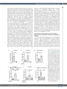
A Bi Bii
Figure 2. Hyaluronic acid treatment increases acute myeloid leukemia cell arrests on VCAM-1 under shear flow. (A) OCI-AML3 cells were per- fused over hyaluronic acid (HA). Where indicated, cells were pretreat- ed with blocking αCD44 antibody (clone 515). (B) OCI-AML3 (i) or pri- mary acute myeloid leukemia (AML) cells from five different patients (ii) were perfused over VCAM-1. Where indicated, cells were pretreated with soluble HA or αCD49d antibody (clone HP2/1) (abrogating VLA-4- mediated interactions). (C) OCI-AML3 cells were perfused over a VCAM-1 or VCAM-1/HA substrate upon pretreat- ment with αCD49d antibody (clone HP2/1), where indicated. (D) OCI- AML3 cells were perfused over VCAM-1. Where indicated, cells were pretreated with low molecular weight HA (LMW-HA) or high molecular weight HA (HMW-HA). (E) OCI-AML3 cells transduced with shCD44 or con- trol shRNA were perfused over VCAM- 1; cells were pretreated with soluble HA, where indicated. Categories of interaction (tethers) are expressed as frequencies of cells in direct con- tact with the substrate. Two groups were compared with a paired t-test, three groups were compared with one-way analysis of variance with multiple comparisons. *P<0.05; **P<0.01; ns: not significant.
CDE
haematologica | 2021; 106(8)
2105
