
Page 17 - 2021_06-Haematologica-web
P. 17
The post-HCT microbiome: effects and targeted therapy
Additionally, features of the intestinal microbiome have been associated with the development of pulmonary infiltrates post-transplant.52 A further study found that the presence of butyrate-producing bacteria in fecal samples was associated with a lower rate of viral lower respirato- ry tract infections.53
Finally, there is also evidence emerging that early post- HCT microbiome damage may remain for some time, and have consequences for the late complications of transplantation. A recent study by our group showed that dysbiosis can still be observed at 100 days after transplan- tation and that patients who go on to develop chronic GvHD have lower concentrations of circulating SCFA.54 As this was a small study of chronic GvHD patients, fur- ther work is needed to explore this association.
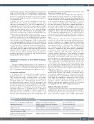
The studies described above found several links between dysbiotic gut microbiota and transplant-related complica- tions but it remains largely unresolved whether these asso- ciations reflect causal relationships. If this is indeed the case, then manipulation of the microbiota – particularly in a fashion that ensures maintenance of anaerobic bacterial taxa and prevents colonization with potential pathogens – could be an effective way to reduce acute transplant-relat- ed toxicities and also improve the long-term outcomes for allogeneic HCT recipients. At present, potential strategies for microbiota modification include dietary modification, prebiotics, probiotics or postbiotics and fecal microbiota transplantation (FMT) (Table 1).
Potential mechanisms of microbiome-targeting therapies
Therapies targeting the microbiota could contribute to preserving or restoring intestinal homeostasis by provid- ing resistance to colonization, enhancing mucosal heal- ing, or dampening the inflammatory immune response.
Colonization resistance
As mentioned above, outgrowth of a single taxon, par- ticularly Enterococcus, is a common feature of the post- HCT microbiome and is associated with a poor trans- plant outcome. In addition to Enterococcus colonization, intestinal expansion of other pathogenic bacteria, such as the Enterobacterales species Klebsiella pneumoniae and Escherichia coli, has been observed in patients who subse- quently developed bloodstream infections caused by the respective species.55 Domination may negatively affect intestinal homeostasis: (i) indirectly via loss of beneficial anaerobic bacteria, resulting in a drastic reduction of ‘health-promoting’ metabolites such as SCFA or, (ii) directly, if the dominating species itself compromises the
gut epithelium or invokes an inflammatory response from the mucosal immune system.
Enterococcus faecalis (for example) has the capacity to impair epithelial barrier integrity via matrix metallopro- teases and can also induce a strong dose-dependent acti- vation of dendritic cells, resulting in enhanced production of the cytokines IL-6, IL-10, IL-12 and TNF-α.56,57 A recent mouse study showed that intake of the disaccharide lac- tose was necessary for Enterococcus expansion whereas a lactose-free diet attenuated the emergence of Enterococcus and reduced the severity of acute GvHD.42 These pre-clin- ical findings need further exploration in clinical trials, for example, of lactose-free diets in the peri-transplant peri- od, or enzymatic supplementation (e.g., with the lactase enzyme (Lactaid), which is cheap and easily available as it is used commonly by individuals with lactose intoler- ance).
Reintroduction of specific strains via defined bacterial consortia in oral capsule form (commonly known as pro- biotics) or transfer of a complete (healthy) microbiome via FMT could prevent domination by a single taxon by promoting resistance to colonization. For example, probi- otic therapy with Lactobacillus johnsonii prevented Enterococcus domination after bone marrow transplanta- tion in mice, which coincided with attenuation of acute GvHD.47 Similarly, FMT effectively eliminated van- comycin-resistant Enterococcus from densely colonized mice, which was mediated by anaerobic bacteria.58 More specifically, recolonization with Barnesiella correlated with eradication of vancomycin-resistant Enterococcus. In these studies, successful engraftment of donor strains was a key component of successful treatment. Factors that could influence this process include pre-treatment strate- gy (i.e., antibiotic decontamination or bowel lavage, with the latter appearing to be more effective), taxonomic con- figuration of both donor and recipient and possibly also genetic host factors.59-61
Alternative approaches that bypass the bacterial com- munity itself but may still enhance colonization resist- ance include supplementation of antimicrobial peptides or administration of specific TLR agonists. For example, TLR7 might be a candidate as activation of TLR7 on den- dritic cells induced IL-22 and mediated colonization resistance against vancomycin-resistant Enterococcus.62 Whether targeting this pathway could also be beneficial for transplant outcome needs further investigation.
Healing of the mucosal barrier
The mucosal barrier becomes compromised during the allogeneic HCT process due to the toxic effects of chemo- and radiotherapy and subsequent mucositis, as a result of tissue destruction during acute GvHD, opportunistic
Table 1. Strategies for targeting the microbiome. Method
Prebiotics (i.e. non-digestible food ingredients)
Optimizing dietary intake
Antibiotic selection
Probiotics/defined consortia (i.e. selected
viable microorganisms) or FMT
Postbiotics (bioactive metabolic compounds)
Strategy
Supplement essential bacterial nutrients
Stimulate enteral feeding, avoid consumption
of certain nutrients
Reduce exposure to anaerobe-targeting antibiotics Actively reintroduce (specific) microbial populations
Goal
Preserve protective taxa
Preserve protective taxa
Limit microbial damage
Recover gut microbial communities
Supplement products of bacterial fermentation
Replenish beneficial microbial metabolites
FMT: fecal microbiota transplantation.
haematologica | 2021; 106(8)
2045
