
Page 147 - 2021_06-Haematologica-web
P. 147
Antibody-mediated procoagulant platelets in VITT
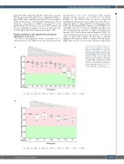
tively, P<0.0001, Figure 5A). On the other hand, sera from VITT patients showed slight but not significant binding to Spike-RBD (Online Supplementary Figure S2D). Most impor- tantly, in the presence of PF4 the IgG binding was reduced when the concentration of RBD is increased above 6.5 mg/mL (Figure 5A). However, sera from VITT patients did not show significant binding to S2 protein with and with- out PF4 (Figure 5B; Online Supplementary Figure S2E).
Platelet activation in the heparin-induced platelet aggregation assay assay
In order to investigate the ability of patients’ sera to activate platelets, the HIPA assay was used with several
A
modifications. Sera were incubated with washed platelets in the presence of i) buffer, ii) 0.2 IU/mL LMWH, iii) 100 IU/mL UFH, iv) an Fcγ receptor IIa (FcγRIIA)-blocking monoclonal antibody (mAb IV.3), v) 30mg/mL IVIG, vi) 25 mg/mL PF4, vii) 50 mg/mL Spike- RBD, viii) PF4/Spike-RBD complexes, ix) PF4+RBD or x) ChAdOx1 nCoV-19. Conditions with PF4 and RBD were also repeated in the presence of high concentration of heparin (100 IU/mL unfractionated heparin [UFH]). We observed platelet activation in the presence of buffer in eight of eight VITT patients (median time to platelet aggregation 5 minutes [min], no range 5-10 min [min*], Figure 6A), but not in sera from vaccinated individuals
Figure 5. Immunoglobulin G binding to SARS-CoV-2 Spike-RBD and S2 in the presence and absence of PF4. (A) Immunoglobulin G (IgG) binding to SARS- CoV-2 Spike-RBD was assessed by IgG- enzyme immune assay (EIA) and expressed as fold increase to PF4 alone. (B) IgG binding to SARS-CoV-2 S2/PF4 complexes was assessed by EIA and expressed as fold increase to PF4 alone. ns: not significant; *P<0.05, **P<0.01, ***P<0.001 and ****P<0.0001.
B
haematologica | 2021; 106(8)
2175
