
Page 145 - 2021_06-Haematologica-web
P. 145
Antibody-mediated procoagulant platelets in VITT
Immunoglobulin G binding profile of sera from vaccine- induced immune thrombotic thrombocytopenia
High-titer PF4/heparin antibodies were detected in all sera (eight of eight, 100%) using the IgG PF4/heparin EIA. Interestingly, binding of all sera was inhibited in the pres- ence of high concentration of heparin (mean optical densi- ty [OD] of IgG antibodies against PF4/heparin complexes: 2.591±0.642 versus 0.176±0.073, respectively, P<0.0001, Figure 3A). No correlation was found between the PF4/heparin antibodies and the detected COVID-19 anti- bodies in VITT patients and in vaccinated controls (Online Supplementary Figure S1A to D [I-IV]). Among non-vaccinat- ed controls only one subject (4%) had a PF4/heparin anti- bodies in EIA (data not shown).
We next investigated the PF4-seroconversion after vacci- nation with ChAdOx1 nCoV-19, as well as during severe SARS-CoV-2 infection (Figure 3B). We found that four of 41 (9.8%) vaccinated healthy individuals and four of 25 (16%) patients with severe COVID-19 seroconverted with IgG antibodies against PF4/heparin complexes within 14 days (Figure 3B). Next, we tested IgG binding to platelets by FC. An increase in IgG binding to test platelets was observed (fold increase [FI] in mean fluorescence [MF]
AB
intensity compared to healthy controls [FI of MFI]: 4.39±1.15 vs. 1±1.10, P=0.026, Figure 4, Online Supplementary Figure S2A). IgG binding to platelets was inhibited by heparin at high concentrations (FI of MFI IgG binding: 1.51±0.66, P=0.016), but not at low concentra- tions (FI of MFI of IgG binding: 3.60±2.01, P=0.688). Only one serum showed increased binding to platelets in the presence of PF4 and the vaccine ChAdOx1 nCoV-19 (case #4, Figure 4). Spike-RBD did not induce a significant change in IgG binding in VITT patients (Figure 5A). Similar results were observed when S2 protein was added (Figure 5B). IgG binding was also observed when sera from ChAdOx1 nCoV-19 vaccinated volunteers with IgG PF4 antibodies were tested (Online Supplementary Figure 2B). However, severe COVID-19 patients with IgG PF4 anti- bodies showed no increase in IgG binding (Online Supplementary Figure S2C).
The impact of Spike-RBD on the binding of anti-PF4 antibodies
Compared to healthy controls, sera from VITT patients showed strong binding to PF4 in the in-house EIA (OD IgG antibodies against PF4: 1.03±0.04 vs. 0.110±0.002, respec-
CDE
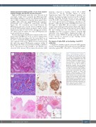
Figure 2. Histopathological findings in case #2 and case #3. (A) Case #2: occlu- sion of glomerular capillary loops by hya- line thrombi. Hematoxylin-Eosin (H&E) staining, original magnification 200x. (B) Deposition of platelets in glomerular ves- sels documented by CD42b staining, immunoperoxidase staining, magnifica- tion 200x. (C) Case #3: occlusion of glomerular capillary loops by hyaline thrombi with fibrin deposits highlighted in red, Masson’s trichrome stain, magnifica- tion 200x. (D) Immunostaining for CD61 and (E) fibrin demonstrate the massive intravascular deposits of fibrin and platelets, immunoperoxidase staining, magnification 200x. (F) Thrombotic occlu- sions of submucosal vessels in the urinary bladder with hemorrhage. H&E staining, magnification 40x. (F) Thrombotic occlu- sion of medium-sized pulmonary vessels. H&E stining, magnification 40x. (G) Insert shows platelet deposits in pulmonary cap- illaries stained for CD61, immunoperoxi- dase staining, magnification 400x.
FG
haematologica | 2021; 106(8)
2173
