
Page 144 - 2021_06-Haematologica-web
P. 144
K. Althaus et al.
bruising and petechiae, which might be an early sign of VITT. One patient underwent a successful thrombus removal by endovascular rheolysis. Three of eight patients died (on day 6 [case #1], day 10 [case #2] and day 7 [case #3] of hospitalization). All surviving patients received anti- coagulation. Four patients received intravenous immunoglobulin (IVIG) combined with non-heparin anti- coagulation.
Pathological findings
Autopsy was performed in two of three deceased patients. Autopsy of case #2 showed complete throm-
botic obstruction of the straight, sagittal and transversal cerebral sinuses, subarachnoidal hemorrhage, cerebral edema and bilateral pulmonary embolism in mid-sized arteries and obstruction of glomerular arterioles and capillaries by hyaline microthrombi containing fibrin and platelets (Figure 2A and B). Autopsy of case #3 showed massive cerebral hemorrhage and cerebral edema, bilateral pulmonary thromboembolism and obstruction of glomeruli by hyaline microthrombi (Figure 2C to G). Histology of the bone marrow was normal in both cases without any hint of increased thrombopoesis.
Table 1. Demographic and clinical data of cases with vaccine induced immune thrombotic thrombocytopenia. Case # Age Sex First symptoms Thrombosis/ Thrombotic PLT, D-Dimer,
9
after vaccination Bleeding risk factors (150-450x10 /L) (<0.5 mg/mL)
(days)
Fibrinogen, INR (170-410 mg/dL)
aPTT, (>40s)
1
2 3
4 5 6 7
8
47 f 7 CVST none 10 >35 128 1.30 23
48 24
53
47
32
36
29
f 6 CVST, PE
m 10 bleeding, multiple
n.a. 40 n.a.
heterozygous 22 n.a. FVL mutation
n.a. 1.16 22.9 109 1.20 42
126 1.01 25
263 1.25 35
n.a. n.a. n.a.
n.a. 1.19 22
274 1.00 23
thrombosis
m 9 DVT,PE none 8 >35
f 7 CVST none 56 9 m 20 PE none 71 n.a. f 17 CVST none 92 13 f 7 CVST contra-ception 53 32
CVST: indicates cerebral venous sinus thrombosis; DVT: deep vein thrombosis; FVL: Factor V Leiden; n.a.: not available; PE, pulmonary embolism; PLT: platelet.
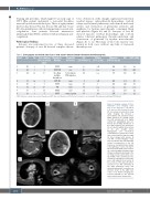
ABC Figure 1. Imaging example of three illustrative cases. Imaging exam- ples of case #1 (A to C), case #8 (D to F) and case #4 (G and H). In case #1, non-enhanced computed tomog- raphy imaging (A) showed a parenchymal and subdural hemor- rhage (arrows in A), causing a mid- line shift (arrowheads in A). Digital subtraction angiography was per- formed (B) showing thrombosis of the right sigmoid and transverse sinus, superior sagittal sinus (arrows in B), and straight sinus. Angiography after mechanical recanalization (C) shows the recanal- ized cerebral sinuses (superior sagit- tal sinus marked with arrows). In case #8, cerebral imaging 7 days after vaccination was unremarkable (curved reconstruction of the left transverse and sigmoid sinus shown in the right upper corner of D and F). She worsened, which led to a repeat- ed cerebral imaging, showing a large intraparenchymal hemorrhage in the left temporal lobe (arrow in E), caus- ing midline shift (arrowhead in E), caused by a thrombosis of the trans- verse and sigmoid sinus (arrows in F), as well as of the adjacent tentori- al veins. In case #4, a thrombus in the right pulmonary artery was observed (arrows in E; coronal recon- struction shown in the right lower corner of G). Further imaging also revealed thrombi in the femoral
DEF
GH
veins on both sides (arrows in H).
2172
haematologica | 2021; 106(8)
