
Page 111 - 2021_06-Haematologica-web
P. 111
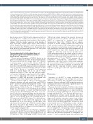
Bile acids reduce GvHD and preserve the GvL effect
Figure 4. Tauroursodeoxycholic acid reduces intestinal antigen presentation. (A to G) Small intestinal samples were isolated from recipient mice treated with vehicle or tauroursodeoxycholic acid (TUDCA), on day 14 after bone marrow transplantation (BMT) (C57BL/6 to BALB/c model). (A) Identification of significantly downregu- lated Gene Ontology terms in animals treated with TUDCA. The dotted line corresponds to P=0.05 (log -0.05=1.29). (B) Heat map based on microarray analysis show- ing the differentially regulated genes (q-value<0.05) that belong to the term ‘antigen processing and presentation’ from the Gene Ontology database. Data were pooled from two independent experiments, n=6 mice per group, P=0.004. The color code represents the Z-score log2 intensity. (C) Quantitative real-time polymerase chain reaction (PCR) analysis of the mRNA expression of selected genes with Actb as a reference gene. Data were pooled from two independent experiments, num- bers (N) represent individual mice. P-values were calculated using the unpaired two-tailed Student’s t-test. (D) Expression of TAP1 protein quantified by western blot. Representative western blot from n=3 mice per group. (E) Quantification of TAP1 protein expression. Data were pooled from two independent experiments, numbers (N) represent individual mice. P-values were calculated using the unpaired two-tailed Student’s t-test. (F) Flow cytometric quantification of major histocompatibility complex (MHC) class I expression on CD326+ (EpCAM+) cells (left panel) and MHC class II expression on CD45– MHC class II+ cells (right panel). Data were pooled from two independent experiments, numbers (N) represent individual mice. P-values were calculated using the unpaired two-tailed Student’s t-test. (G) Heat map based on microarray analysis showing the differentially regulated genes (q-value<0.05) that belong to the term ‘Response to interferon γ’ from the Gene Ontology database. Data were pooled from two independent experiments, n=6 mice per group, P=6.49x10⁻⁹. The color code represents the Z-score log2 intensity. (H and I) Quantitative real-time PCR analysis of the mRNA expression of Tap1 (panel H) and Tap2 (panel I) with Actb as a reference gene in MODE-K cells treated with TNF ± chenodeoxycholic acid (CDCA), ursodeoxycholic acid (UDCA), 6-ethylchenodeoxycholic acid (obeticholic acid, OCA) and TUDCA for 48 hours. Representative data from one of two independent experiments with n=3 replicates/group are presented. P-values were calculated using the ordinary one-way ANOVA test with correction for multiple comparisons; ns: not significant.
had shown a role for TUDCA and its chaperone activity for reducing ER stress, a cellular stress response to critical con- ditions that can potentially lead to apoptosis. We found no changes of ER stress marker expression in the intestine of mice developing aGvHD when treated with TUDCA (Online Supplementary Figure S6C and D). Altogether, these data demonstrate that TUDCA protects the intestinal tract from apoptosis and particularly preserves ISC and goblet cells from aGvHD-related damage.
Tauroursodeoxycholic acid treatment does not abrogate the graft-versus-leukemia activity or hematopoietic regeneration
We next assessed the impact of TUDCA directly on the graft-versus-leukemia (GvL) effect. Reduced antigen presen- tation and human leukocyte antigen (HLA) loss contribute to immune escape of acute myeloid leukemia (AML) cells after allo-HCT.25 In order to test whether bile acid treat- ment alters the MHC/TAP antigen presentation in leukemic cells, we treated four human and murine leukemia cell lines with TUDCA. HLA-A, B, C and MHC class I expression were not altered (Figure 7A, B, D and E; Online Supplementary Figure S7). Also Tap1 and Tap2 gene expres- sion remained unchanged, suggesting that bile acid applica- tion does not impair the GvL activity by reducing the expression of MHC/TAP molecules on malignant cells (Figure 7C and F; Online Supplementary Figure S7).
We then activated CD8+ T cells by co-incubation with allogeneic dendritic cells and treated them with TUDCA prior to incubation with the A20 lymphoma cell line. T cells were capable of inducing cell death in the leukemic cells which remained stable in the case of TUDCA addition (Figure 7G). We tested in vivo T-cell priming by re-isolating T cells from the spleens of vehicle- and TUDCA-treated animals 14 days after BMT (Figure 7H). These in vivo acti- vated T cells had a comparable killing capacity when incu- bated with A20 cells (Figure 7I). Finally, we studied the GvL effect in vivo by injecting Ba/F3 cells containing the FLT3- ITD translocation (Ba/F3-ITD) (Figure 7J). Additional trans- fer of alloreactive T cells reduced the expansion of the malignant cells in the bone marrow and the spleen. This effect persisted in the mice upon TUDCA treatment (Figure 7K). These data indicate that TUDCA affects specifically the intestine and does not impede cytotoxic lymphocyte activity against malignant cells.
Since immune reconstitution is critical for a favorable outcome after allo-HCT, we investigated whether TUDCA might have a negative impact on the peripheral blood cell reconstitution. Mice developing aGvHD treated with
TUDCA and vehicle displayed the expected decrease in hemoglobin, hematocrit, platelets and white blood cells (WBC) in comparison to untreated mice (Online Supplementary Figure S8A). TUDCA did not enhance cytopenia as treated animals exhibited the same blood counts as vehicle controls. WBC subpopulation analysis by flow cytometry revealed similar proportions of T cells, B cells, granulocytes and monocytes in the peripheral blood (Online Supplementary Figure S8B). Analysis of mice with aGvHD at a later time point was not possible due to GvHD-induced mortality in the vehicle group. In order to segregate aGvHD from immune reconstitution, we ana- lyzed animals transplanted only with BM without a GvHD- inducing transfer of T cells. TUDCA treatment did not compromise hematopoietic regeneration (Online Supplementary Figure S8C and D). These results underline the observation that TUDCA treatment does not prolong the time period necessary for immune reconstitution and suggest that this compound would not impair immune responses to pathogens or the GvL response in allo-HCT patients.
Discussion
Frequency of allo-HCT is rising worldwide since improved conditioning protocols and supportive care allow older patients to undergo this potentially curative leukemia treatment. However, efficient GvHD prophylaxis remains a challenge in the clinical routine with a significant impact on the long-term outcome of allo-HCT.
Here, we observed a depletion of the bile acid pool after allo-HCT. We hypothesized that loss of bile acids might be one of the factors contributing to loss of homeostasis in the intestinal tissue during GvHD (Figure 8). In agreement with recently published data,6,7 we observed that exposure to pro-inflammatory cytokines caused intestinal tissue dam- age in an organoid culture model. Treatment with bile acids improved the viability of intestinal organoids and cell lines when cell death was induced by application of TNF or IFNγ. The role of bile acids in the regulation of cell death has been controversially discussed.26 Depending on their concentra- tion, they can have cytotoxic properties and are able to induce apoptosis either by direct activation of death recep- tors, for example FAS, or by inducing oxidative damage and mitochondrial dysfunction.27-29 Other studies indicate that there is a fine balance in bile acid composition in order for their toxic and protective properties to antagonize one another.30,31 The secondary hydrophilic bile acid UDCA has
haematologica | 2021; 106(8)
2139
