
Page 109 - 2021_06-Haematologica-web
P. 109
Bile acids reduce GvHD and preserve the GvL effect
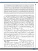
Figure 3. Bile acid treatment improves acute graft-versus-host disease outcome in mice. (A) Transplantation model with BALB/c (H-2kd) as donor and C57BL/6 (H- 2kb) as recipient. Recipient animals were treated with 200 mg/kg body weight tauroursodeoxycholic acid (TUDCA) or an equal volume of vehicle from day 0 until day 10 after bone marrow transplantation (BMT) by a daily intraperitoneal injection. (B) Survival of C57BL/6 mice transplanted as shown in (A). Numbers (N) represent individual mice, the P-value was calculated using the two-sided Mantel-Cox test. (C) Transplantation model with C57BL/6 (H-2kb) as donor and BALB/c (H-2kd) as recipient. Recipient animals were treated with 200 mg/kg body weight TUDCA or an equal volume of vehicle from day 0 until day 10 after BMT by a daily intraperi- toneal injection. (D) Survival of BALB/c mice transplanted as described in (C). Data were pooled from three independent experiments, numbers (N) represent indi- vidual mice. The P-value was calculated using the two-sided Mantel-Cox test. (E) Graft-versus-host disease (GvHD) histopathology scores of liver, small intestine and colon assessed on day 7 after BMT (C57BL/6 in BALB/c model). Data were pooled from two independent experiments, numbers (N) represent individual mice. P-values were calculated using the ordinary one-way ANOVA test with correction for multiple comparisons. (F) Serum cytokine concentrations in untreated mice and transplanted mice determined on day 14 after BMT (C57BL/6 in BALB/c model). Numbers (N) represent individual mice. P-values were calculated using the ordinary one-way ANOVA test with correction for multiple comparisons. (G and I) Fecal samples were collected for microbial analysis on day 5 after BMT. Numbers (N) represent individual mice. Data were pooled from two independent experiments. P-values were calculated using the ordinary one-way ANOVA test with correction for multiple comparisons; ns: not significant. (G) The Shannon index as a surrogate parameter for microbial diversity. (H) The reversed Simpson index as a surrogate parameter for microbial diversity. (I) Relative abundance of specified bacterial genera. (J) C57BL/6 mice were treated with an antibiotic cocktail comprising 1 mg/mL cefoxitin, metronidazole, neomycin and gentamycin for 2 weeks before they underwent BMT as described in (A). (K) Survival of C57BL/6 mice transplanted and treated as described in (J). Numbers (N) represent individual mice. Data were pooled from two independent experiments. Statistical analysis was performed using the two-sided Mantel-Cox test.
to E). Downregulated genes included transporter associated with antigen processing 1 and 2 (Tap1 and Tap2) (Figure 4B to E), which are involved in the translocation of degraded cytosolic peptides across the endoplasmatic reticulum membrane for antigen-major histocompatibility complex (MHC) class I molecule assembly, as well as TAP binding protein (Tapbp) and Tapasin-related protein (Tapbpl) which mediate the association between TAP proteins and newly assembled MHC class I complexes. Furthermore, lower transcription of class II MHC complex transactivator (Ciita) was observed suggesting decreased MHC class II-related antigen presentation in the intestines of TUDCA-treated animals (Figure 4B and C). Confirming the hypothesis that the antigen presentation machinery in the intestine is reduced, we found decreased levels of MHC class I and MHC class II expression on non-hematopoietic cells in the intestine of TUDCA-treated animals (Figure 4F). In line with data obtained from the organoid culture system, mul- tiple genes associated with the GO term ‘Response to IFNγ’ were significantly downregulated (Figure 4G). Interestingly, the numbers of CD11c+ MHC class II+ professional antigen- presenting cells (APC) in ileum and colon were identical between both groups (Online Supplementary Figure S4A). There was a slight reduction in TNF expression, whereas costimulatory molecules and other cytokines remained unchanged (Online Supplementary Figure S4B and C). The migration capacity of bone marrow-derived dendritic cells (BM-DC) was not impaired by TUDCA treatment either (Online Supplementary Figure S4D). These data support the hypothesis that antigen presentation by non-hematopoietic cells is reduced by TUDCA treatment. In line with this model, treatment with TNF elevated the expression of Tap1 and Tap2 in MODE-K cells which could be reversed by addition of TUDCA (Figure 4H and I). Notably, the effect on Tap1/2 expression was most evident for TUDCA but occurred upon treatment with CDCA and UDCA as well (Figure 4H and I).
Bile acid administration changes the transcriptional signature of T cells in the intestine but preserves their systemic expansion
Antigen presentation is important for the recognition of malignant cells by alloreactive T cells. Since TUDCA reduces antigen presentation in the intestine, we asked whether its application has an immediate impact on T-cell activation. T-cell numbers in the lamina propria of the small intestine and production of IFNγ, interleukin-6 (IL-6) and TNF were not altered upon TUDCA administration (Figure 5A; Online Supplementary Figure S5A and B). However, we
discovered a transcriptional signature suggesting downreg- ulation of immune cell activation. We observed that the transcriptional levels of many genes associated with the GO term ‘T-cell activation’ were significantly reduced in GvHD developing mice, treated with TUDCA (Figure 5C). Among these genes were the CD3 subunits Cd3e and Cd3g, the transcription factor interferon regulatory factor 1 (Irf1), as well as many genes encoding for proteins that play an important role for signaling pathways downstream of the T-cell receptor (Figure 5D). Among these were lympho- cyte-specific protein tyrosine kinase (Lck) and linker for acti- vation of T cells (Lat). Lck is a tyrosine kinase that phospho- rylates the tails of the CD3 chains of the T-cell receptor complex upon antigen recognition via MHC. This allows ZAP70 binding and activation. Lat is phosphorylated by ZAP70/Syk kinases upon T-cell receptor activation and recruits adapter proteins which are important for further signaling. This altered transcriptional signature suggests that multiple events in the downstream signaling of the CD3/T-cell receptor complex are reduced upon treatment with TUDCA.
We then investigated whether TUDCA administration could decrease the general alloreactive T-cell expansion. Bioluminescence-based trafficking analysis revealed that T- cell expansion was similar in vehicle- and TUDCA-treated animals (Figure 5E to G). Flow cytometry confirmed that there were no differences in T-cell numbers in the spleen between both groups (Online Supplementary Figure S5C to F). T-cell differentiation, migration and TNF production after in vitro stimulation and TUDCA treatment were com- parable (Online Supplementary Figure S5G to I). Collectively, our results support the hypothesis that bile acid application is also associated with a transcriptional signature of reduced local T-cell activation without having a negative impact on systemic T-cell expansion.
Tauroursodeoxycholic acid decreases apoptosis in the intestine
We observed that bile acids prevent intestinal cell death in vitro. Further analysis of the microarray data obtained from the in vivo allo-HCT model revealed a significant downreg- ulation of apoptosis-related genes in mice treated with TUDCA (Figure 6A). Confirming this, we performed an immunofluorescent TdT-mediated dUTP-biotin nick end labeling (TUNEL) staining which marks apoptotic nuclei. While aGvHD induction significantly increased intestinal apoptosis, TUDCA administration reduced it to almost the baseline level in both the small intestine and the colon (Figure 6B and C). The application of TUDCA increased the
haematologica | 2021; 106(8)
2137
