
Page 49 - 2021_07-Haematologica-web
P. 49
Synergistic targeting of WEE1 and GLS1 in T-ALL
WEE1 expression in different T-ALL subtypes from 264 primary T-ALL samples, based on unique gene expression signatures reflecting distinct stages of arrest during T-cell development.32 Interestingly, WEE1 mRNA levels varied with the highest expression in the TLX1 subtype (Figure 1D). Regardless of these variations, we identified a global upregulation of WEE1 in T-ALL.
MYC directly activates WEE1 transcription in T-cell acute lymphoblastic leukemia
To understand the molecular mechanism underlying WEE1 upregulation in T-ALL, we conducted in silico analy- sis in the UCSC genome browser gateway to identify potential transcription factor regulating WEE1 expression. MYC, TAL1 and GATA3 were predicted to activate the WEE1 promoter (Online Supplementary Figure S2A). We
individually knocked down each of these transcription factors, using two individual shRNA (sh#1 or sh#2) in human T-ALL cells, and found that only depletion of MYC decreased WEE1 mRNA and protein levels (Figure 2A and Online Supplementary Figure S2B). Consistently, bromod- omain 4 (Brd4) inhibitor JQ1, which represses MYC tran- scription,33 decreased WEE1 mRNA and protein levels con- comitant with downregulation of MYC expression in multiple T-ALL cells (Figure 2B and Online Supplementary Figure S2C). In contrast, JQ1 treatment, inhibiting N-MYC as well,34 caused minimal effect on the WEE1 steady-state level in T-ALL LOUCY cells which predominantly express N-MYC (Online Supplementary Figure S2D). In addition, inactivation of MYC similarly down-regulated WEE1 (expression in Burkitts lymphoma P493 cells (Online Supplementary Figure S2E). These data suggest that MYC,
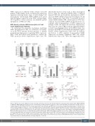
AB
CDEF
GH
Figure 2. MYC directly activates WEE1 transcription. (A) Jurkat and CUTLL1 cells were infected with lentiviruses expressing control (shCtrl) or MYC shRNA (shMYC#1 or shMYC#2). WEE1 mRNA and protein levels were determined by real-time-quantitative polymerase chain reaction (RT-qPCR) and immunoblots. ACTIN served as a loading control. (B) CCRF-CEM and KOPTK1 cells were treated with JQ1 as indicated, and WEE1 protein levels were determined by immunoblots. (C) Schematic pres- entation of MYC binding site (E-box, -39 ~ -34) on the WEE1 promoter. The potential MYC responsive element (wild-type, WT) and its mutant (MUT) are shown as indi- cated. (D) Binding of MYC to the WEE1 promoter was analyzed by chromatin immunoprecipitation (ChIP) in CUTLL1 cells. Averages of fold enrichment between MYC and isotype IgG are shown. NCL was analyzed as a positive control. (E) Luciferase reporter activities of the WEE1 promoter (-210−342bp) containing MYC RE-WT (and MUT) were detected in the presence of ectopically expressed MYC in 293T cells. 3xMYC E-box (3Ebox) sequences were used as a positive control. Reporter activities relative to empty pGL3-Basic vector (Vector) are shown. (F) Correlation of WEE1 expression with MYC in 117 primary T-cell acute lymphoblastic leukemia (T-ALL) sam- ples (GSE26713). (G) Correlation of WEE1 expression with MYC in 264 primary T-ALL samples. (H) Correlation of WEE1 expression with MYC in NOTCH1 mutant or
2
WT T-ALL cases shown in (G). Gene expression levels from primary T-ALL samples are presented in Log represent the means (± standard deviation) of biological triplicates. **P<0.01.
scale. Data of RT-qPCR and luciferase reporter analysis shown
haematologica | 2021; 106(7)
1819
