
Page 241 - 2021_06-Haematologica-web
P. 241
Letters to the Editor
ABC
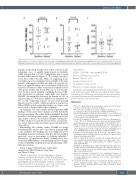
Figure 2. Effect of chelation on pituitary iron and volume. A longitudinal study (mean 2.6 years) showed a significant decrease in pituitary iron (P<0.02) corre- lating with an improvement in cardiac iron (P<0.03). The change in pituitary volume was minimal, as expected to be irreversible, and not statistically significant.
patients, respectively. Results were stable or worse in the remaining ones. A parallel improvement in pituitary Z(R2) was noted in 9/17 (53%) individuals, whose mean baseline Z(R2) was 6.4 (Figure 2). The change was mod- est in those with a baseline Z(R2) >8, suggesting chela- tion failure in severe pituitary iron load. Eight of the nine patients with improved Z(R2) also had a decrease in car- diac iron, again showing a good correlation of these two measures. Pituitary volume remained abnormally low in subsequent testing. The mean Z(V) was -2.6 at baseline and -2.4 upon repeat imaging, confirming the irreversible cell destruction in patients with high iron burden. LH/FSH concentrations decreased by a mean of 28% at repeat testing in 7/8 women. AMH levels remained sta- ble overall, supporting reports on preserved gonadal function and successful ovulation induction despite low LH and FSH levels. Longitudinal studies on a larger scale are needed to further explore these findings.
In summary, our data confirm previous pituitary MRI findings of iron and volume changes and risk thresholds for hypogonadism. Our study further highlights the high prevalence and progressive nature of pituitary iron load- ing, despite advances in chelation therapy, causing irre- versible volume loss and deleterious effects on reproduc- tive status. Splenectomy and older age are risk factors for these effects.
Our findings also validate cardiac T2*MRI as a proxy for pituitary R2, which can be used where pituitary MRI is not available. The findings also have implications for modifying chelation treatment, which is effective in mild- moderate cases in preventing or delaying infertility.16 Discussion of reproductive status, utilizing these hor- monal and MRI findings, and informing patients of high- er risk of infertility are prudent for proper planning of fer- tility preservation.
Sylvia T. Singer,1 Roland Fischer,2 Isabel Allen,3 Ashutosh Lal,1 Elliott Vichinsky,1 Qing Yuan4
and Zhiyue J. Wang4
1Division of Hematology-Oncology, Department of Pediatrics, University of California San Francisco, UCSF Benioff Children’s Hospital Oakland, Oakland, CA, USA; 2University Medical Center Hamburg-Eppendorf (UKE), Hamburg, Germany; 3University of California San Francisco, San Francisco, CA, USA and 4University of Texas Southwestern Medical Center, Texas, TX, USA
Correspondence:
SYLVIA T. SINGER - sylvia.singer@UCSF.edu doi:10.3324/haematol.2020.252726
Received: March 23, 2020.
Accepted: October 9, 2020.
Pre-published: October 29, 2020.
Disclosures: no conflicts of interest to disclose.
Contributions: STS designed and performed the study, analyzed results and wrote the paper; ZJW and QY analyzed patients' images and pituitary data; RF reviewed and analyzed the data and reviewed and revised the manuscript; IEA performed the statistical analysis; AL and EV reviewed and revised the paper.
References
1. Wood JC. Estimating tissue iron burden: current status and future prospects. Br J Haematol. 2015;170(1):15-28.
2. Wood JC. Use of magnetic resonance imaging to monitor iron over- load. Hematol Oncol Clin North Am. 2014;28(4):747-764, vii.
3. Noetzli LJ, Panigrahy A, Hyderi A, Dongelyan A, Coates TD, Wood JC. Pituitary iron and volume imaging in healthy controls. AJNR Am J Neuroradiol. 2012;33(2):259-265.
4. Noetzli LJ, Panigrahy A, Mittelman SD, Hyderi A, Dongelyan A, Coates TD, et al. Pituitary iron and volume predict hypogonadism in transfusional iron overload. Am J Hematol. 2012;87(2):167-171.
5. Wang ZJ, Wang DJ, Chia JM, Yuan Q, Morriss MC, Rollins NK. A software tool for semi-automated quantification of pituitary vol- umes. International society of magnetic resonance imaging in med- icine (ISMRM) 19th meeting. Montreal, Canada, 2011:4247.
6. La Marca A, Broekmans FJ, Volpe A, Fauser BC, Macklon NS. Anti- Mullerian hormone (AMH): what do we still need to know? Hum Reprod. 2009;24(9):2264-2275.
7. Yalti S, Gurbuz B, Ficicioglu C. Serum levels of inhibin B in men and their relationship with gonadal hormones, testicular volume, testic- ular biopsy results and sperm parameters. J Obstet Gynaecol. 2002;22(6):649-654.
8.Aydinok Y, Bayraktaroglu S, Yildiz D, Alper H. Myocardial iron loading in patients with thalassemia major in Turkey and the poten- tial role of splenectomy in myocardial siderosis. J Pediatr Hematol Oncol. 2011;33(5):374-378.
9.Poggi M, Sorrentino F, Pugliese P, et al. Longitudinal changes of endocrine and bone disease in adults with beta-thalassemia major receiving different iron chelators over 5 years. Ann Hematol. 2016;95(5):757-763.
10. Casale M, Citarella S, Filosa A, et al. Endocrine function and bone disease during long-term chelation therapy with deferasirox in patients with beta-thalassemia major. Am J Hematol. 2014; 89(12):1102-1106.
haematologica | 2021; 106(6)
1743
