
Page 219 - 2021_06-Haematologica-web
P. 219
Hypoxia signature in BIA-ALCL
roring their cellular expression levels (Figure 6A). Since BIA-ALCL cells can secrete CA9, we next examined whether CA9 could be detected in peri-implant seroma specimens involved by BIA-ALCL. Indeed, all ten BIA- ALCL seroma specimens evaluated contained detectable CA9, with a mean concentration of 84,046 ± 118,695 pg/mL (range, 423-360,262 pg/mL) (Figure 6B). In contrast, seromas lacking involvement by BIA-ALCL showed a mean concentration of 502 ± 390 pg/mL (range, 9-887 pg/mL; P<0.0001, Mann-Whitney test).
Four serum and/or plasma samples from BIA-ALCL were available to evaluate CA9 concentrations (Online Supplementary Figure S3). While the CA9 concentration in normal human serum or plasma is <25 pg/mL,24,26 the plas- ma CA9 concentration was 128 pg/mL in one BIA-ALCL patient. Because the number of human blood samples available for testing was limited, we examined whether CA9 secreted from BIA-ALCL cells might be detectable in serum samples using mouse xenograft models. We har- vested subcutaneous TLBR-1, -2, and -3 tumors when each tumor reached a volume of 1,000 mm3 and obtained simultaneous serum samples. Tumor lysate CA9 concen- trations from TLBR-1, -2, and -3 were 108,175±39,252 pg/mL, 231,070 ± 88,185 pg/mL, and 6,903 ± 1,871 pg/mL, respectively, based on standardized total protein concen- trations of 1 mg/mL; all pairwise comparisons showed sig- nificant differences (Online Supplementary Figure S4). A similar pattern of serum CA9 concentrations was observed, with mean values for TLBR-1, -2, and -3 tumor- bearing mice of 170 ± 46 pg/mL, 183 ± 170 pg/mL, and 122 ± 119 pg/mL; values in all groups were significantly higher than CA9 concentrations in serum obtained from non-tumor-bearing mice (46 ± 11 pg/mL) (Figure 6C). Taken together, these findings indicate that CA9 can be secreted from BIA-ALCL cells and is detectable in peri- implant seroma fluid involved by BIA-ALCL. Serum CA9 concentrations are elevated in sera from BIA-ALCL xenograft-bearing mice and serum levels in BIA-ALCL patients should be evaluated in larger cohorts.
notable findings, including upregulation of genes involved in cell motility (e.g., CCR6, MET, and HGF), myeloid cell differentiation (e.g., PPARG and JAK2), and viral gene tran- scription (e.g., RPS10), and downregulation of T-cell receptor signaling genes. Differentially expressed genes reported by Di Napoli et al. tended to show similar changes in our dataset (Online Supplementary Figure S6). However, the gene ontology analysis of Di Napoli et al. compared BIA-ALCL to non-neoplastic T cells, whereas our study was designed to identify differences between
A
Discussion
In this gene expression profiling study comparing BIA- ALCL to their non-BIA counterparts, we found that BIA- ALCL demonstrate a hypoxia signature, likely attributable to the unique microenvironment in which they arise. Notably, the carbonic anhydrase CA9 was expressed con- sistently in BIA-ALCL and only minimally in non-BIA- ALCL. CA9 promoted hypoxia-induced growth in BIA- ALCL cell lines in vitro and in mouse xenograft models. In addition, CA9 was significantly elevated in human seroma samples involved by BIA-ALCL and in serum from BIA- ALCL xenograft-bearing mice. These findings identify unique biological features of BIA-ALCL, support its classi- fication as a WHO entity distinct from other forms of ALCL, and uncover opportunities to explore hypoxia-relat- ed proteins and pathways in novel diagnostic, preventive, or therapeutic strategies for patients with this disease.
RNA sequencing with transcriptomic analysis and GSEA revealed enhanced expression of hypoxia signaling pathway genes as a hallmark of BIA-ALCL. A recent study by Di Napoli et al. also compared the transcriptome of BIA-ALCL to that of other peripheral T-cell lymphomas including non-ALCL.27 The authors identified a number of
C
B
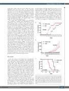
Figure 5. CA9 accelerates breast implant-associated anaplastic large cell lym- phoma growth in a mouse xenograft model. (A) Mice inoculated with TLBR-3 breast implant-associated anaplastic large cell lymphoma cells stably trans- duced with a CA9 lentiviral vector develop palpable tumors faster than mice inoculated with cells transduced with vector control. (B) Mice inoculated with TLBR-3 cells overexpressing CA9 develop larger tumors than mice inoculated with control-transduced TLBR-3 cells. (C) Overall survival is shorter in mice bear- ing CA9-overexpressing TLBR-3 tumors than in those bearing control-transduced tumors.
haematologica | 2021; 106(6)
1721
