
Page 220 - 2021_06-Haematologica-web
P. 220
N. Oishi et al.
BIA-ALCL and TN ALCL arising at other anatomic sites. Therefore, these two studies are complementary and emphasize different aspects of BIA-ALCL pathogenesis for further study. Of note, occasional non-BIA-ALCL (especially with TN genetics) showed a moderate degree of CA9 expression, highlighting the need for additional future study of hypoxia-associated pathways in T-cell neoplasms other than BIA-ALCL.28
BIA-ALCL arises in a unique tumor microenvironment consisting of the breast prosthesis, seroma fluid, and sur- rounding fibrous capsule. Local hypoxia is a well-estab- lished factor promoting the development of tissue fibro- sis,29,30 and tissues with artificial prostheses are postulated to be hypoxic.31,32 For example, Kim et al. showed that the thickness of the fibrous capsule around silicone implants in rats was reduced by stem cell-derived endothelial precur- sor cell conditioned medium, which promotes wound healing at least in part by reducing tissue ischemia, suggest- ing the peri-implant microenvironment is hypoxic even in the non-neoplastic setting.33 The ability to tolerate low oxygen tension may be critical for pre-neoplastic cells situ- ated between the prosthesis and peri-implant fibrous cap- sule to survive and proliferate in the early stages of BIA- ALCL lymphomagenesis. Since most patients with implants do not develop BIA-ALCL, however, future stud- ies should examine possible interplay between hypoxia and recurrent genetic events reported in this disease, such as mutations in JAK-STAT and epigenetic modifier genes.17 Furthermore, it would be of interest to compare the molec- ular signature of BIA-ALCL with that of other effusion- associated malignancies such as primary effusion lym- phomas of B-cell origin, in which targetable hypoxic meta- bolic pathways have been reported previously.34,35
Among genes within the hypoxic signature, we identi- fied CA9 as being most robustly overexpressed in BIA- ALCL, a finding we validated at the protein level by immunohistochemistry. CA9 is a hypoxia-inducible enzyme that catalyzes reversible hydration of carbon diox- ide to bicarbonate ions and protons.22 CA9 is expressed in a variety of solid cancers and has been associated with poor prognosis.36-38 Overexpression of CA9 represents an adaptive response to hypoxia by which cancer cells control intracellular and extracellular pH, facilitating survival and growth in an acidic tumor microenvironment.22,39-44 Our data on silencing CA9 expression in hypoxia-inducible TLBR-2 cells and overexpressing CA9 in hypoxia-insensi- tive TLBR-3 cells indicate that CA9 promotes growth of BIA-ALCL cells. Although CA9 inhibitors have been devel- oped,23 the direct therapeutic implications of our findings for BIA-ALCL are unclear since disease limited to the hypoxic seroma and surrounding capsule is adequately managed by surgery alone in most cases.3 We did not have adequate tissue material from disseminated BIA-ALCL to evaluate whether CA9 expression is retained outside its native microenvironment. Nevertheless, understanding the role of CA9 and other hypoxic signaling pathways in early BIA-ALCL could lead to less invasive strategies to manage localized disease and/or novel prosthetic approaches that decrease the risk of its development.
Our findings also suggest that CA9 could be useful as a biomarker for screening, detection, and/or follow-up of BIA-ALCL. CA9 expression in normal human tissues is limited to gastric, colonic, and gallbladder epithelium.22 Clear cell renal cell carcinoma is a prototypic malignancy expressing high CA9, in which recurrent VHL mutations
lead to increased expression of hypoxia-associated genes including CA9; accordingly, CA9 is a widely-used immunohistochemical marker to distinguish clear cell renal cell carcinoma from other renal tumors.45 In addition, serum CA9 levels are associated with tumor size, grade,
A
B
C
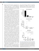
Figure 6. CA9 as a candidate biomarker in breast implant-associated anaplas- tic large cell lymphoma. (A) TLBR-1, -2, and -3 cell lines secrete CA9 into culture supernatant proportionally to their cellular expression, as determined by west- ern blot (cf. Figure 3A). Data represent three replicates measured by CA9 enzyme-linked immunosorbent assay. (C) Peri-implant seroma samples involved by breast implant-associated (BIA) anaplastic large cell lymphoma (ALCL) have significantly higher CA9 concentrations than those not involved by BIA-ALCL. (C) Serum samples obtained from mice bearing 1000 mm3 subcutaneous TLBR-1, - 2, and -3 tumors have significantly higher CA9 concentrations than those from non-tumor-bearing mice. *P<0.05; **P<0.01; ***P<0.001; ****P<0.0001 (Mann-Whitney test). PBS: phosphate-buffered saline.
1722
haematologica | 2021; 106(6)
