
Page 186 - 2021_06-Haematologica-web
P. 186
R.D. Jachimowicz et al.
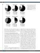
Figure 2. Cellular composition assessed by whole-slide imaging in the study cohort and according to histological subtypes of classi- cal Hodgkin lymphoma. Average values of percentage of the respective cell types are indicated. For calculation of cell counts of macrophages (CD68) and Hodgkin and Reed-Sternberg cells (CD30) see the Online Supplementary Methods.
which express CD73 (Online Supplementary Figure S4). Thus, the vast majority of B cells seem to resemble naïve B cells. However, further subtyping of B cells in the microenvironment using appropriate methods is required to understand the nature of this population.
Inter-observer bias of whole-slide image analysis
To evaluate the inter-dependent bias of WSI, we took a two-pronged approach. First, we randomly selected CD20- stained whole slides that were re-analyzed by a second independent observer, using the same software. We found a high inter-observer concordance when the same or newly scanned images were processed in the image analy- sis software by a second observer (Online Supplementary Figure S6). Second, we randomly selected a cohort of 20 cases, for which the complete staining procedure for CD20 and WSI was performed in a second center (see Online Supplementary Methods). This approach revealed a correla- tion of results obtained at two independent centers (r2=0.6101) (Online Supplementary Figure S7). Thus, WSI provides an unbiased approach for robust quantification of the global cellular composition of cHL tissue.
Discussion
Despite the overall outstanding treatment results in cHL, the a priori identification of a high-risk subset of patients remains a challenge in clinical practice. Studies
utilizing functional imaging influence clinical decision- making,22 but none of the previously proposed gene expression profiling or immunohistochemistry biomark- ers has been incorporated into treatment protocols for cHL.4 So far, a comprehensive analysis of the cellular com- position of the microenvironment in cHL is limited to gene expression studies.12,16,23 Here, we utilized WSI and achieved a comprehensive and robust quantification of the cellular composition of the cHL microenvironment throughout the whole tumor sample. To the best of our knowledge, this approach has not been applied to cHL previously and opens a novel conceptual window into the assessment of cellular composition of tumor tissue.
In contrast to previously reported studies,11,17,24 but in line with several other studies,25,26 we did not observe a corre- lation of macrophage content with outcome in cHL. The discrepancy between our results and previously published studies with respect to macrophage counts may be explained in multiple ways. First, the technology to assess macrophage counts differs. We cannot rule out that gene expression analysis utilizing mRNA expression level of multiple genes assesses macrophages in a different man- ner than our WSI approach. However, comparing conven- tional immunohistochemistry image analysis or even visu- al inspection of small fields of view, we consider WSI a more accurate measure of macrophage content. Second, there is a difference between the population of patients in our study and those in previous publications. Patients in our study were treated with rather intensive chemothera- py (BEACOPP/eBEACOPP), which is known to achieve
1688
haematologica | 2021; 106(6)
