
Page 188 - 2021_06-Haematologica-web
P. 188
R.D. Jachimowicz et al.
A
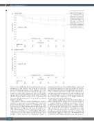
Figure 3. Progression-free sur- vival according to CD20 con- tent. (A) Kaplan-Meier plots of progression-free survival (PFS) in the study cohort for the two risk groups according to B-cell content (CD20-positive cell rate: ≤21% or >21%). (B) Kaplan-Meier plots of PFS in the validation cohort for the two risk groups according to B- cell content (CD20-positive cell rate: ≤10% or >10%).
B
types of tumor-infiltrating B cells populating the microen- vironment of solid cancers,32 the adoption of a broader panel of B-cell/plasma-cell-associated antigens, T-cell sub- sets and correlation with PD-1 staining should be per- formed in future WSI applications and could include, among others, CD4, CD8, CD19, CD27, CD5, CD38 and CD138. However, WSI on large cohorts, as performed in our study, will probably have to be focused on a limited number of markers.
Nevertheless, WSI has several advantageous features, compared to gene expression profiling and conventional immunohistochemistry studies, by combining the diag- nostic accuracy of digital image analysis and a large-scale approach. By providing cell counts (e.g., the number of B cells) independently of relative expression levels of bio- markers for cell subtypes (e.g., CD20 mRNA), WSI reflects a direct read-out for the cellular composition and conse- quently a direct measure for therapeutic targets of
immunological therapy. Since CD20 staining is performed at virtually any diagnosis of cHL, WSI makes use of exist- ing histology data without any additional molecular pro- cedures. CD20-stained slides of cHL can be digitalized at any pathology center and moved via the worldwide web in a timely manner to allow centralized assessment. We thus envision this technology to be highly suitable for incorporation into future clinical trials.
The cellular composition of the microenvironment of cHL is complex and its analysis has so far been restricted to a limited number of lymphoma specimens.33 WSI of multiplexed-stains allows the number of cellular markers to be increased and might help in the translation of find- ings obtained in a few patients’ specimens into large cohorts of patients. Moreover, novel analytic tools beyond the plain assessment of relative amount of cell types can potentially be applied to WSI data.34 In summary, B-cell content assessed by WSI in advanced-stage cHL allows for
1690
haematologica | 2021; 106(6)
