
Page 172 - 2021_06-Haematologica-web
P. 172
T. Suzuki et al.
A
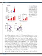
Figure 2. Strong correlation between bone marrow PPARd mRNA and mobilization efficacy. (A) Alteration of PPAR family mRNA in bone marrow (BM) cells during granulo- cyte colony-stimulating factor (G-CSF) mobi- lization (n=3-5). (B, C) Stepwise increase along with increasing G-CSF doses in (B) PPARd mRNA in BM cells and (C) colony- forming units in culture (CFU-C) in the blood (n=3-5). (D, E) Correlation of mobilization efficiency of CFU-C with (D) white blood cell (WBC) in the blood and (E) PPARd mRNA in BM cells in mice fed with a normal diet (ND, violet dots; n=22) or a fat-free diet (FFD, orange dots; n=4). ns: not significant. Combined data from at least three inde- pendent experiments are shown. Data are mean±standard error of mean. *P<0.05, **P<0.01, ***P<0.001 (Student t test, analysis of variance, and Pearson correla- tion coefficient).
BC
DE
(Figure 2A). The increase in the expression of BM PPARd and mobilized HPC in the blood was dependent on the number of G-CSF doses (Figure 2B and C). Based on these data, we analyzed the correlation between mobilization efficiency and BM PPARd mRNA expression in a subset of C57BL/6 male mice fed with a ND after eight doses of G- CSF. The number of mobilized CFU-C varied greatly (range, 1200-3900/mL blood), and white blood cell count showed only a correlation trend (Figure 2D). Although the correlation between mobilized LSK cells and BM PPARd mRNA was weak and not statistisically significant (Online Supplementary Figure S3), mobilization efficiency by CFU- C correlated strongly with BM PPARd mRNA (Figure 2E, violet dots). We also performed the same analysis in a sub- set of mice fed with the FFD. Consistent with this corre- lation, both mobilization efficiency and BM PPARd mRNA were higher than those of the best mobilizer mice fed the ND (Figure 2E, orange dots). Thus, in G-CSF mobi- lization, higher expression of BM PPARd is itself a marker of better mobilization. More importantly, this higher mobilization efficiency was likely due to the lack of sig- naling of this fatty acid ligand-activated transcription fac- tor as a result of the insufficient supply of fat in the BM.
Next, we tried to identify the cell types that express
PPARd protein in BM. Immunohistochemical staining revealed clearly increased PPARd expression after eight doses of G-CSF treatment. Morphologically, myeloid line- age cells, which were relatively large with various seg- mental shaped nuclei, were positive, whereas small round lymphocytes with little cytoplasm were negative for PPARd (Figure 3A). PPARd protein and mRNA expression was also evaluated in sorted myeloid cell fractions. Flow cytometric analysis revealed that all three major myeloid populations in the BM, i.e., mature neutrophils (CD11b+Ly6GhighF4/80low) immature neutrophils (CD11b+Ly6GdullF4/80low) and monocytes/macrophages (CD11b+Ly6GdullF4/80high), showed high expression in steady-state, and both mature and immature neutrophils displayed a significant increase in PPARd protein and mRNA following G-CSF treatment (Figure 3B–D). In con- trast, PPARd protein expression in these three myeloid fractions in peripheral blood was observed in only minor populations and it was not increased by G-CSF treatment (Online Supplementary Figure S4), indicating the marrow- specific role of PPARd.
Next, the alteration of BM PPARd mRNA expression by the depletion of mature neutrophils was examined using the anti-Ly6G antibody, 1A8 (Online Supplementary Figures
1674
haematologica | 2021; 106(6)
