
Page 171 - 2021_06-Haematologica-web
P. 171
Mobilization regulated by PPARd
Results
A short-term fat-free diet enhances mobilization efficiency
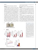
To examine the effect of insufficient fat intake on mobi- lization, WT C57BL/6 male mice were fed with a FFD, containing sufficient calories with protein and all known vitamins but without fat, or a ND for 2 weeks. The body weight of mice fed with the FFD for 2 weeks was compa- rable to that of the mice fed with the ND (24.71±0.32 and 24.14±0.35 g, respectively; n=11). The administration of either G-CSF (8 divided doses, every 12 h, 125 mg/kg/dose, s.c.) or vehicle (PBS/bovine serum albumin [BSA]) was fol- lowed after this period with the continuation of the same diet. This period of fat restriction was reported to be safe with regard to sequelae associated with deficiency of essential fatty acids.19,20 This simple regimen had a dramat- ic effect, with the number of hematopoietic progenitor cells (HPC) being increased in the circulation compared to that in mice fed with a ND, as assessed by lineage-Sca-1+c- kit+ (LSK) cells and colony-forming units in culture (CFU- C) (Figure 1A and B; Online Supplementary Figure S1A) with no alteration in BM HPC (Online Supplementary Figure S1B). Enhanced mobilization was also confirmed in hematopoietic stem cells (HSC), as assessed by long-term competitive repopulation for 6 months (Figure 1C). Thus,
A
B
a short-term deficit in fat intake is a promising method to enhance mobilization efficiency.
Marrow PPARd expression correlates with mobilization efficiency
Enhanced mobilization was unlikely due to the alter- ation of known key players in BM microenvironment for mobilization, such as osteolineage cell activity and a chemokine, because mRNA levels of Runx2, osteocalcin, and CXCL12 in BM cells after G-CSF treatment were comparable between animals fed the FFD or the ND (Online Supplementary Figure S2). According to this obser- vation, we hypothesized that some lipid mediators from food intake might play a role in the BM to inhibit mobi- lization, and that a FFD led to a lack of these BM lipid mediators.
We first searched for a possible receptor that could induce this inhibitory signal. The PPAR family consists of fatty acid ligand-activated transcription factors.21 Among all three PPAR, α, γ, and d (b/d), in BM cells, PPARα mRNA was unchanged. Consistent with the previously reported suppression of PPARγ in CXCL12-abundant reticular cells by G-CSF,22 PPARγ mRNA was significantly suppressed after G-CSF treatment (Figure 2A). Meanwhile, PPARd mRNA displayed the highest expression in the steady state and increased dramatically after G-CSF mobilization
Figure 1. Short-term fat restriction enhanced hematopoietic stem/progenitor cell mobilization by granulocyte colony- stimulating factor. (A) Macrophotographs of culture dishes (35 mm) showing the results of the colony-forming units in cul- ture (CFU-C) assay for mobilization of cells into the peripheral blood (20 mL) following eight doses of granulocyte colony-stimulat- ing factor (G-CSF). ND: normal diet, FFD: fat-free diet. (B) Mobilization efficiency assessed by the presence of white blood cells (WBC), lineage-Sca-1+c-kit+ (LSK) cells, and CFU-C in the blood (n=4 for group treat- ed with phosphate-buffered saline (PBS)/bovine serum albumin (BSA) and n=10-11 for the group treated with G-CSF). (C) Hematopoietic stem cell activity assessed by a long-term competitive repop- ulating assay of mobilized blood. Repopulating units (RU) were evaluated at 6 months after competitive transplantation (n=6). Representative pictures or com- bined data from at least three independent experiments are shown. Data are mean ± standard error of mean. *P<0.05, **P<0.01, ***P<0.001 (Student t test and Mann-Whitney U test).
C
haematologica | 2021; 106(6)
1673
